Download
1 / 28
290 likes | 476 Views
Head Injury (HI). Hassan Bukhari General and Trauma Surgeon Apr 29, 2012. Objective. By the end of this discussion, you should be familiar with Anatomy and physiology of the cranium Classification of HI Diagnosis Clinical Imaging. Contents. Introduction Classification of HI Diagnosis

E N D
Head Injury (HI) Hassan Bukhari General and Trauma Surgeon Apr 29, 2012
Objective • By the end of this discussion, you should be familiar with • Anatomy and physiology of the cranium • Classification of HI • Diagnosis • Clinical • Imaging
Contents • Introduction • Classification of HI • Diagnosis • Treatment • MCQs
Introduction • Trauma is the leading cause of death in young patients worldwide • Head injury (HI) accounts for >50% • Goals of Intervention in HI • To reduce mortality and improve outcome • To prevent secondary brain damage • Cannot do much about primary brain damage
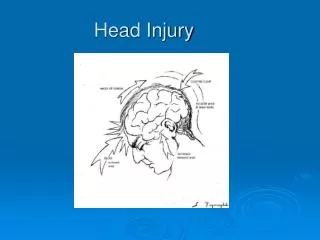
Anatomy of Cranium • SCALP • Skull • Meninges • Brain • Ventricles • Tentorium
Classification • Mechanism • Blunt vs penetrating • Severity • Minor • Moderate • Sever • Morphology • Skull fracture • Intracranial lesions
Intracranial lesions • Focal • Epidural • Subdural • Subarachnoid • Intracerebral • Diffuse • Concussion • Diffuse axonal injury • Multiple contusions
Epidural hematoma (EH) • Convex collection • More in young • Source: MMA • Incidence: about 10% • MVC, fall and assault
Epidural hematoma • Lucid interval: observed in up to 50%. • About 40% will remain conscious throughout. • Mortality • Overall is 10%
Subdural hematoma • Extracranial,crescentic collection • Source: bridging vein • MVC, fall and assault
Diagnosis • Clinical
Diagnosis • Imaging • Skull x-ray • CT • MRI
MCQ #1 • 34 YO M, involved in MCV, present in coma. CT brain as shown, Diagnosis is A- Subdural B- Epidural C- Subarachnoid D- Intracerebral bleed E- Brain contusion ✓
MCQ #2 • A 44 YO, fall from 5 meters. Present with CGS of 8. CT as shown. Diagnosis is: A- Subdural B- Epidural C- Subarachnoid D- Intracerebral bleed E- Brain contusion ✓
MCQ #3 • 57 YO F, was hit by baseball bat to the head. She had transient loss of consciousness. On arrival, she is full conscious and asymptomatic. During observation, she became confused and her GCS is 10. What do you call this phenomena? A- Lucid phase interval B- Cushion interval C- Rebound phenomena D- Countercoup phenomena ✓
Treatment • Medical • General • Specific • Conservative • Control ICP • Surgical • Decompressive craniotomy
MCQ #4 • A 20 YO M, involved in MCC. Came to ER with the following finding (see photo). These finding are A- Raccoon eyes B- Rhinorrhea C- Basal skull fracture D- All of the above E- Non of the above ✓
In summary • Anatomy and physiology of the cranium • Classification of HI • Diagnosis • Clinical • Imaging
Reference • American Association of Neurological Surgeons: Guidelines for Surgical Management of TBI 2006. Neurosurgery 58:S2-1-S2-3, 2006. • American College of Surgeons: ATLS® Student Course. 8th edition