Download
1 / 75
840 likes | 1.35k Views
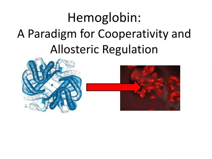
Hemoglobin: A Paradigm for Cooperativity and Allosteric Regulation. Why do we breathe?. http://www.uni.edu/schneidj/webquests/spring04/tvbroadcast/circulatorysystem.html. Cellular Requirement for O 2. Oxygen Carriers. Diffusion Limited solubility of O 2 in Blood and Cell Water.

E N D
Hemoglobin:A Paradigm for Cooperativity and Allosteric Regulation
Why do we breathe? http://www.uni.edu/schneidj/webquests/spring04/tvbroadcast/circulatorysystem.html
Oxygen Carriers Diffusion Limited solubility of O2 in Blood and Cell Water
Myoglobin and Hemoglobin • Myoglobin (Mb) • Increases O2 solubility in tissues (muscle) • Facilitates O2 diffusion • Stores O2 in tissues • Hemoglobin (Hb) • Transports O2 from lungs to peripheral tissues (erythrocytes)
Function(s) of Myoglobin Facilitate O2 Diffusion in Muscle O2 Storage (aquatic mammals)
Structure of Sperm Whale Myoglobin Figure 7-1
The Heme Prosthetic Group Figure 7-2
Properties of Heme Prosthetic Group in Myoglobin • Tightly bound • Synthesized separately from myoglobin • Fe2+ Coordination • Nitrogens of heme (4) • His (F8): proximal histidine • His (E7): distal histidine • Ligands: O2, CO, and NO
Ligands Small molecules that bind to proteins by non-covalent interactions (e.g. O2 to myoglobin)
Ligand Binding • usually transient and reversible interaction with others molecule (= ligands) such as metals, hormones • often involves “molecular breathing” of the protein, i.e. ability to undergo small conformational changes • often induces molecular rearrangements in the protein • ligand binding sites are • - highly conserved • - complementary in size, shape, and charge
Heme • prosthetic (permanent, non-proteinaceous) • groupof Mb and Hb • incorporated into Hb and Mb during folding • responsible for reversible O2 binding • responsible for red color of blood and muscles
Heme – Structure central Fe2+ 4 methyl groups 2 vinyl groups (buriedin protein) 2 propionate groups(exposed)
Heme – Iron Coordination • Fe2+ has 6 coordination sites • 4 with N of pyrrole rings, • 2 perpendicular to ring • Mb/Hb: 5th coordination site is occupied with proximal His • 6th coordination site: • O2 oxyhemoglobin • none deoxyhemoglobin • CO carboxyhemoglobin
Heme – Binding of CO vs. O2 • free heme binds C0 105 times better than O2 • kinked binding topology in Mb/Hbfavors O2 (100-fold) • TOTAL: CO binding ~ 230 fold stronger than O2 binding (Carbon monoxide poisoning)
Function(s) of Myoglobin Facilitate O2 Diffusion in Muscle O2 Storage (aquatic mammals)
Myoglobin (Mb) • primarily found in muscle (highly abundant in marine mammals such as whales) • single polypeptide (153 aa) with one bound heme • very simple oxygen binder: binds oxygen at high pO2, releases it at low pO2 • Mb + O2 MbO2 • typical globin fold
The Globin Fold 8 helices (A-H) and loops in between MCDB310 – Chapter 5: Protein Function
Binding/Association Constant Ka Quantitatively describes the affinity of a protein P for its ligand L P + L PL the higher the binding affinity, the higher Ka
Dissociation Constant Kd P + L PL the higher the binding affinity, the smaller Kd Example: Ka = 106 M-1Kd = 10-6 M
Degree of Saturation, Fraction of binding sites that are occupied by ligand at any given ligand concentration 0 1
Degree of Saturation, Using If [L] = Kd = 0.5 Kd is the ligand concentration at which 50% of the binding sites are occupied
Some Examples with KD = 1 µM Question: What fraction of the protein has ligand bound when the [L] is 1 µM or 10 µM? [L] = 1 µM: [L] = 10 µM:
Myoglobin – Oxygen Binding Curve Revisited When ligand is a gas, partial pressures = concentrations
Myoglobin – Oxygen Binding Curve Revisited • Saturation of Mb depends on • the binding constant of Mb for O2 (KD = p50 = 2.8 torr) • the concentration of O2 (pO2) • Question: What is the fractional saturation of Mb? • pO2 = 1 torr: • pO2 = 10 torr:
Myoglobin – An Oxygen Storage! pO2 in lung ~ 13 kPa pO2 in tissue ~ 4 kPa 10 kPa = 76 torr
Hemoglobin (Hb) • present in erythrocytes (makes blood look red, 34% of weight is Hb) • Different Hb subtypes: • Hb A (adult): two (141 aa) and two (146 aa) subunits that are arranged as a pair of identical subunits (2 subunits) • Hb F (fetal): two and two chains
Hemoglobin – 3D Structure a2 b1 a1 b2
Each subunit has 1 heme, which binds 1 O2 O2 Heme Lehninger, Figure 7-5, 7-6
Hemoglobin • Erythrocytes: • 1 ml blood: 5 x 10⁹ erythrocytes • 1 erythrocyte: 3 x 10⁸ Hb molecules • Hb is a good marker for number of red blood cells • Homology: • 50% of AA are identical between and subunits • 20% of AA are identical between / and Mb
Function of Hemoglobin O2 binding in lungs O2 release in tissues
Oxygen binds to hemoglobin and myoglobin differently Myoglobin Hemoglobin
Oxygen binding to hemoglobin pO2 = partial pressure of oxygen Θ = fraction of binding sites that are occupied
p50 is the pO2 where half the binding sites are occupied p50
Hb has evolved to transport O2 pO2 In Tissues pO2 In Lungs 38% p50
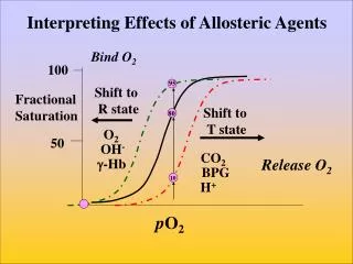
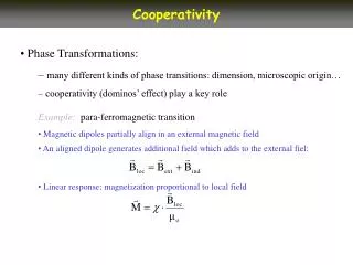
Hb gains cooperativity by switching between 2 states T state (Low Affinity) R state (high affinity) Lehninger Figure 7-10
The Concerted Model All or nothing mechanism T R Lehninger, Figure 7-14
The Concerted Model All or nothing mechanism T R Lehninger, Figure 7-14
Hb follows a little of both T R Lehninger, Figure 7-14
Movements of the Heme and the F Helix During the T —> R Transition Figure 7-8
Local structural changes around the Heme are communicated to the rest of Hb By Janet Iwasa, https://iwasa.hms.harvard.edu/project_pages/hemoglobin/hemoglobin.html
Changes in the 1–2 Interface during the T —> R Transition in Hemoglobin Figure 7-9