Download
1 / 33
340 likes | 889 Views
Diverticular Disease . Dr. Matt W. Johnson. Introduction & Overview. Pathology Physiology Location Complications Bleeding Obstruction Fistula Acute Diverticulitis Management of Acute Diverticulitis. Pathology. Congenital Acquired

E N D
Diverticular Disease Dr. Matt W. Johnson
Introduction & Overview • Pathology • Physiology • Location • Complications • Bleeding • Obstruction • Fistula • Acute Diverticulitis • Management of Acute Diverticulitis
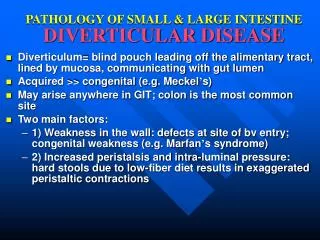
Pathology • Congenital • Acquired • association with Western diets high in refined carbohydrates and low in dietary fibre1 • Deficiency of vegetable fibre in diet2 • Disordered motility • Hyperelastosis may lead to structure change • Collagen abnormalities • Age • Diverticular disease occurs in over 25% of the population, increasing with age3 1 Ferzoco et al Lancet 1998; 2 Simpson et al Br J Surg 2002; 3 Janes et al BJS 2005
Physiology • La Place effects • High intra-luminal pressure • Resultant characteristic protrusion mucosa • Worst at terminal arterial branches • Rectal sparing • ?due to complete layer of longitudinal muscle and large diameter
Physiology and Anatomy • Terminal arterial branches • Penetrate circular muscle • Often lie adjacent to taenia
Location • Classically Sigmoid • In Orient often right-sided • Rectal Sparing • Can occur anywhere(but considered separately)e.g. Small bowel –see later
Specimen showing blood in diverticulae Complications • Obstruction • Bleeding • Inflammation “itis” • Fistula • Sepsis • Perforation • May co-exist with IBD
Obstruction in Diverticular Disease • Progressive distension • Single contrast enema will delineate this • Often present like cancer • Diagnosis • often only at operation (opened specimen) or • on histology
Bleeding in Diverticular Disease • Rarely exsanguinating • Often requires repeat transfusion • Consider mesenteric angiography if available • Embolisation (risk of ischaemia and infarction) • Allows targeted resection • Operative intervention uncommon • On table colonoscopy • Exclusion
Re-Bleeding Rates Re-bleeding rate Year Percentage 1 9 2 10 3 19 4 25 1 Longstreth Am J Gastro 1997
Other Causes Of Colonic Bleeding • Exclude • IBD • Neoplasm • Angiodysplasia • Ischaemic colitis • Radiation proctitis • Varices
Fistula • Abnormal connection • Commonest communications are • Colovesical • Colovaginal (esp if prev TAH) • Colovesical Symptoms • Pneumaturia • Recurrent infections • Faecalent urine or particulates • Diagnosis of site/communication vs pathology • CD/CRC/TCC
Acute Diverticulitis • Abscess • Peridiverticular • Mesenteric • Pericolic • Perforation • Concealed • Free • Peritonitis (gangrenous sigmoididits) • Purulent or serous or faecal • Local or generalised or pelvic 1 Killingback Surg Clin North Am 1983
Emergency Presentation • Symptoms • Generally unwell • Pain localising to left iliac fossa* • Abdominal distension • Altered bowel habit e.g. diarrhoea • Nausea/Fever • Signs • LIF tenderness • *Beware RIF pain-in right sided diverticulitis and where sigmoid crosses midline • Systemic signs (T/HR/BP/WCC) • May be palpable on pR at anterior rectal wall
Management • Resuscitation • Analgesia • Bloods • ECG/Catheter/Urine • Rectal examination (+/-sigmoidoscopy) • CXR • AXR • USS • CT Scan • Operative intervention
CT Scan Perforated diverticulitis of the sigmoid colon-CT
Operative considerations • Serial assessment and clinical judgement • (even if Radiological perforation) • Operative indications • generalized peritonitis • uncontrolled sepsis, • visceral perforation • acute clinical deterioration • At operation • Resection better than no resection1 • Hartmann’s vs anastomosis 1 Krukowski & Matheson Br J Surg 1984
Anastomosis • Is there any role for primary anastomosis in the inflamed bowel? • Consider if fully resuscitated and colorectal Surgeon • Retrograde gun/washout kit • Schilling et al. 2001 Diseases of the Colon and Rectum • diverticulitis with peritonitis • 13 patients one stage • 42 Hartmann’s procedure • 7% mortality in both groups • Similar complication rates • Not a study of bowel obstruction
Elective Presentation • Via outpatients • Often milder version of emergency presentation • Incidental radiological finding • AXR • Contrast study e.g. Barium Enema • CT scan • Rarely if insiduous, an abscess may be found on Barium Enema as an outpatient
Elective resection for Diverticultis • After recovering from an episode of diverticulitis the individual risk of an urgent Hartmann’s is 1 in 2000 patient-years of follow-up. • Surgery for diverticular disease has a high complication rate • 25% of patients have ongoing symptoms after bowel resection (IBS/IBD) • No evidence to support the idea that elective surgery should follow two attacks of diverticulitis. • Further prospective trials are required. 1 Janes et al BJS 2005
Duodenal and Jejunal Diverticulosis • Separate from colonic diverticulosis. • Most occur in the jejunum and occasionally duodenum. • Jejunal diverticula are acquired protrusions of the mucosal lining through the muscular wall of the bowel. • Encourages particular bacterial overgrowth. • A combination of alteration of the intraluminal contents by these bacteria may result in malabsorption • Calcium • Iron • Vitamins D or B12. • Patients may present with anaemia and occasionally osteomalacia.
Questions ??