Download
1 / 25
250 likes | 401 Views
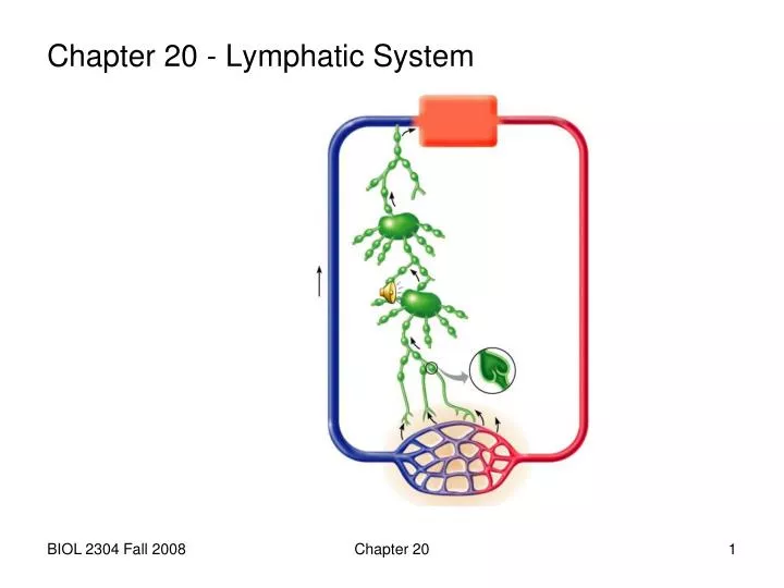
Chapter 20 - Lymphatic System. functions of lymphatic system: prevents edema by removing extra fluid and proteins from the tissues and returning them to the blood immune surveillance

E N D
Chapter 20 - Lymphatic System Chapter 20
functions of lymphatic system: • prevents edema by removing extra fluid and proteins from the tissues and returning them to the blood • immune surveillance cells located in the lymph nodes monitor lymph for pathogens and cancer cells and initiate immune responses if any are detected Chapter 20
the lymphatic system consists of: • various sizes of lymphaticvessels that pick up and transport fluid • lymph, the fluid that is being transported • lymphoidtissues and organs that are located along the lymphatic vessels and that help provide immunity to disease Chapter 20
A. lymphatic vessels (p. 596) • 1. lymph capillaries • smallest lymph vessels • found in loose c.t. near vascular capillaries • (except in the CNS • or bone marrow) Chapter 20
the capillary wall is made of a single layer of endothelial cells that overlap and are very loosely connected • the capillaries are blind-ended (unlike vascular capillaries that have two ends: in and out) Chapter 20
fluid enters lymph capillaries when tissue fluid pressure is high • the overlapping cells let the fluid go in but not back out • pathogens and cancer cells hanging around in the tissues also enter lymph capillaries and are transported to the blood (as they pass through lymph nodes they “alert” the immune system to their presence) Chapter 20
2. lymphatic collecting vessels • each one “collects” lymph from many lymphatic capillaries • found beside veins and arteries • walls similar to veins but thinner • have many valves that prevent backflow 3. lymph trunks • each collects lymph from several collecting vessels • each drains a large area of the body (example: jugular trunks drain neck and head) Chapter 20
4. lymph ducts = largest lymphatic vesselssee diagram in textbook on page 597 and 599 • a. right lymphatic duct drains right arm, head and neck • enters the junction of the right internal jugular and subclavian veins • b. thoracic duct drains rest of body • enters the junction of the left internal jugular and subclavian veins Chapter 20
meet “lymph guy” (p. 597) • he shows you the • parts of the body • drained by the • lymphatic ducts • green-right duct • tan-thoracic duct Chapter 20
B. lymphoid organs (lymph nodes, spleen, MALT) • lymphoid tissue functions: • activation of lymphocytes immunocompetent B and T lymphocytes spread to lymphoid tissue and wait until the foreign antigen they can recognize activates them and starts an immune response • generation of memory cells once a B or T cell is activated, it clones (makes millions of identical copies); some of the clones fight the infection and most die off, but some stick around and wait in case the same foreign antigen gets into the body again—these are called memory cells Chapter 20
lymphoid tissue is a special type of c.t. containing: • reticular cells (fibroblasts) and reticular fibers that form the framework of the organ • T and B lymphocytes that are immunocompetent • macrophages all lymphoid tissue contains lymphatic follicles (also called lymphatic nodules) Chapter 20
a lymphatic follicle (nodule) • is a cluster of lymphocytes derived from a single activated B cell • has a lighter-staining germinal center, the zone where mitosis is occurring • the cloned B cells • differentiate into plasma • cells and leave the • nodule to make • antibodies • (p. 598) Chapter 20
1. lymph node or gland (p. 597) • a. location • clusters are found in some areas of the body and all along lymphatic collecting vessels Chapter 20
b. structure (p. 598) • 1 to 25 mm in diameter • surrounded by fibrous capsule which extends inwards as trabeculae • hilus = indentation on one side • outer cortex contains follicles or nodules • inner medulla contains medullary cords (B and T cells) Chapter 20
afferent lymphatic vessels enter on convex side • inside the node, lymph flows through lymph sinuses (large lymphatic capillaries) and leaves through: • efferent lymphatic vessels exit at hilus Chapter 20
c. functions of a lymph node • monitor lymph • destroy infectious microorganisms and cancer cells • store memory cells Chapter 20
2. spleen (p. 605, 607) located posterior and lateral to the stomach outer fibrous capsule splenic artery branches into central arteries that enter the spleen at the hilus Chapter 20
white pulp = lymphoid tissue that surrounds arteries, destroys blood-borne antigens red pulp surrounds white pulp and contains venous sinuses and splenic cords (reticular tissue rich in macrophages that remove old blood cells) other function: to store platelets Chapter 20
3. MALT - mucosa associated lymphoid tissue • aggregated lymph nodules found in mucosa of the respiratory, digestive, urinary and reproductive tracts Chapter 20
a. tonsils (p. 608, 616) • consist of lymphatic tissue in the mucosa of the pharynx • surface covered by epithelium that invaginates to form crypts that trap bacteria • generate memory lymphocytes Chapter 20
pharyngeal tonsil is in the • nasopharynx on the back wall • tubal tonsils surround openings • of auditory tubes in nasopharynx • palatine tonsils are on the lateral • sides of the opening from the • mouth into the pharynx • lingual tonsils are on the posterior • surface of the tongue Chapter 20
b. GALT (gut) (p. 609) • Peyer's patches in ileum • vermiform appendix • c. BALT (bronchioles) Chapter 20
C. thymus (p. 606) • does not contain lymphoid tissue or B cells • 1. location • posterior to sternum • superior to heart • 2. structure • 2 lobes, each divided into smaller lobules • cortex generates antigen specific T lymphocytes • medulla involved in immune tolerance (destroys T cells that would attack normal body cells) Chapter 20
epithelial reticular cells secrete thymic hormones (thymosin and thymopoietin) that cause T cells to become immunocompetent • most active during childhood • begins to atrophy in adolescence • replaced by fibrous and fatty tissue • protected by a thymus-blood barrier that keeps antigens out cortex medulla Chapter 20
3. function • programs lymphocytes to become immunocompetent T cells • has no direct immune function Chapter 20