Download
1 / 30
680 likes | 1.91k Views
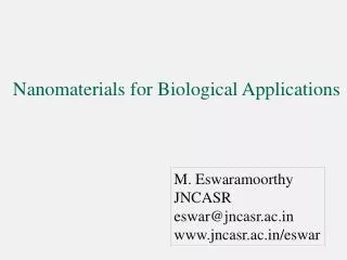
Nanomaterials for Biological Applications. M. Eswaramoorthy JNCASR eswar@jncasr.ac.in www.jncasr.ac.in/eswar. 3D. 1D. 0D. 2D. Ultrathin film. Nanorod, tube. A Nanomaterial has at least one of its dimensions in the nanometric regime. A block of metal. Clusters, Nanoparticles.

E N D
Nanomaterials for Biological Applications M. Eswaramoorthy JNCASR eswar@jncasr.ac.in www.jncasr.ac.in/eswar
3D 1D 0D 2D Ultrathin film Nanorod, tube A Nanomaterial has at least one of its dimensions in the nanometric regime • A block of metal Clusters, Nanoparticles Missing dimension falls in the ‘Quantum Regime’!
Usually exhibit characteristic physical and chemical properties different from the bulk as a consequence of having at least one spatial dimension in the size range of 1-1000 nm.
Examples of Nanomaterials Size (dia.) Materials Nanocrystal and clusters Other nanoparticles Nanowires Nanotubes Nanoporous solids 2-D array of nanoparticles 3-D structures(superlattices) Surfaces and thin films 1-10 nm 1-100 nm 1-100 nm 1-100 nm 0.5-10 nm nm2 - µm2 Several nm in 3-D thickness 1-1000 nm Metals, semiconductors, magnetic materials Ceramic oxides Metals, semiconductors, oxides, sulfides Carbon, layered metal chalcogenides Zeolites, phosphates Metals, semiconductors, magnetic materials Metals, semiconductors, magnetic materials A variety of materials
At the Nanoscale….. • Properties of metals and semi-conductors (electronic, optical, thermodynamic, magnetic) are in between bulk and atoms/molecules. • Size dependent • Surface area is very high • Surface atoms to bulk ratio is more
At the Nanoscale… • Metal can become non-metal! • Hg at 1-2 nm level shows non-metallic band-gap (300 atoms are needed to close the gap) Even the inert gold become very active catalyst at 2 nm size. The color also changes from gold to red, purple etc., • Melting temp. of 2 nm CdS is around 800 ºC and for bulk CdS is around 1700 ºC
Colour is associated with collective oscillation of electrons in the conduction band. -Surface Plasmon Resonance absorption
Quantum Dots CdSe Quantum Dots in hexane • Origin of color due to quantum confinement of electrons • Smaller the quantum dot, larger the band gap, higher the exciton absorption and emission frequency. • Colourtuned with particle size! http://web.mit.edu/chemistry/nanocluster/home.html
Abalone shells Mollusc shell
Inner region- aragonite Outer layer -calcite Prismatic layer Nacreous layer 50µm 1µm Mollusc shell made up of both calcite and aragonite- spatially separated in distinct part of the shell!
In nacreous layer, the aragonite polygonal tablets(500 nm thick) sandwiched between thin sheets(30 nm thickness) of protein-polysaccharide matrix. • It reduces the number of voids in the shell wall and so inhibits propagation of cracks by dissipating the energy along the organic layers rather than through the inorganic crystals. • Nacre 3000 times as tough as inorganic aragonite!!
A: Sheet-like B: Globular C: fibrillar TEM image of silica nanoparticles present in some cell wall of the leaves
A tongue to scar! 7 cm long. Goethite crystals, α-FeOOH (~50 nm) 600 nm Limpets and chitons (sea urchin) have unusual dining habits. Scraping the algae off the intertidal rocks using this radula.
Crystal morphologies and intracellular organization of magnetite nanoparticles inside magnetotactic bacteria: a) cubooctahedral b) bullet-shaped c-d) pseudohexaganol Bars are 100 nm. E. Bauerlein, AngewChemieInt Ed, 2003
Ferrihydrite (5Fe2O3.9H2O) ~5 nm 20 nm Cross-section of ferritin molecule with polypeptide shell and mineral core Discrete nanoparticles of ferritin Acts as a buffer in regulating the levels of free iron – Protects the cells from the pathology of iron overload.
Nanomaterials in Biology Imaging -MRI -Optical Drug Delivery Systems -Meoporous silica -liposomes -nanocapsules Biosensing -Colorimetric -Flouroscence Medicine -Hyperthermia -Labelling -Multiplexed assays
Liposomes Liposomes are soft drug delivery systems loaded with drug in their interiors. Liposomes have the ability to fuse with the cell membrane to deliver the drug in to the cell
Porous nanoparticles (MCM 41) For drug delivery
Drug Delivery Systems Mesoporous silica Nanoparticles Photo induced Release
Fe3O4 @ Silica For MRI and Photothermal Fe3O4 @ Au J. Shi et al, Adv. Funct. Mat. 2008 N. J. Halas et al., Nano Lett. 2006
Photothermal cancer treatment by Au nanocages Au cages shows strong SPR absorption in NIR region where the absorption by blood and tissue is minimal. Large absorption cross-section facilitates the Conversion of NIR absorption into heat SKBR-3 breast cancer cells treated with immuno (anti-HER2)-Au nanocages and then irradiated by 810 nm laser for 5 min. Shows a well-defined circular zone of dead cell c) Calcein AM assay and d)Ethidium homodimer-1(EthD-1). e, f) Without immuno-Au cages Green colour Living cells Red colour- Dead cells Xia et al, JACS 2006 & Warsen et al, Nano Lett, 2007
Semiconductor nanoparticles Metal nanoparticles For imaging and colorimetric sensors, photothermal treatment Susie Eustis and Mostafa A. El-Sayed, Chem Soc. Rev, 35, 209, 2006 http://web.mit.edu/chemistry/nanocluster/home.html
IMAGING Optical Imaging MRI- Magnetic Resonance imaging Magnetic nanoparticles are used as MRI contrast reagents. BRET- bioluminescent resonance energy transfer Brain showing tumor Nature Biotechnology 24, 339 - 343 (2006)
Companies involved in nanoparticle commercialization Salata, J Nanobiotechnology. 2004; 2: 3
Nanomaterials in the pharmaceutical pipeline Dendrimers Drug delivery, imaging agents VivaGel (SPL 7013) Fullerenes Drug delivery, therapeutics Antioxidant fullerene Lipsomes Drug delivery Doxil Superparamagnetic Drug delivery, imaging Ferumoxitol particles agents Ferumoxtran-10 , , Colloidal gold particles Drug delivery, therapeutics Aurimune, Aurothol Polymeric Nanoparticles Drug delivery, therapeutics, Basulin,Contramid imaging MRX-951 , Gold nanoshells Photo-thermal tumour ablation AuroLase Quantum dots Imaging, diagnostics QDot-800 conjugate Self-assembling peptide/protein particles Drug delivery, therapeutics Abraxane Marina A. Dobrovolskaia & Scott E. McNeil Nature Nanotech, vol2, 469, 2007