Download
1 / 14
140 likes | 305 Views
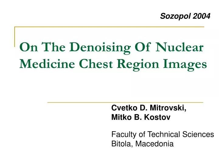
Sozopol 2004. On The Denoising Of Nuclear Medicine Chest Region Images. Faculty of Technical Sciences Bitola, Macedonia. Cvetko D. Mitrovski, Mitko B. Kostov. Structure. Aim / Problem formulation NM images creation process Wavelet shrinkage The filtration of images Experimental results

E N D
Sozopol 2004 On The Denoising Of Nuclear Medicine Chest Region Images Faculty of Technical Sciences Bitola, Macedonia Cvetko D. Mitrovski, Mitko B. Kostov
Structure • Aim / Problem formulation • NM images creation process • Wavelet shrinkage • The filtration of images • Experimental results • Conclusion
Aim of the Work & Problem Formulation • AIM: To develop methods for analyzing of anatomical data and ROIs on a basis of a raw NM image (set of raw NM images). • PROBLEM: To find a suitable method for automatic preprocessing of the chest region NM images & extraction of the anatomical data.
NM Images Creation Process • The raw NM images are based directly on the total counts • a low signal-to-noise ratio (SNR) • noisy due to low count levels, scatter, attenuation, and electronic noises in the detector/camera • One of the major sources of error is Poisson noise due to the quantum nature of the photon detection process
Wavelet Shrinkage Program • DWT (produces two groups of coefficients with low and high SNR) • = wihi • hihard = • hisoft = • Inverse wavelet transformation
Filtration of Chest Region Images • Wavelet shrinkage (threshold for Poisson model?) • the Anscombe variance-stabilizing transformation: • the Donoho’s level dependent threshold: • give up the perfect reconstruction (QMF bank – near PR) Poisson Gaussian noise model
The Algorithm • transformation of the image • calculation of Donoho’s threshold (s =MAD/0.6745) MAD is the median of the magnitudes of all the coefficients at the finest decomposition scale • wavelet soft-thresholding • inverse wavelet transform • square the result • removing shadow in the obtained image
The QMF Bank QMF bank has overall reconstruction error minimized in the minimax sense; the corresponding QMF filters have least-squares stopband error
Comparison with biorthogonal wavelets
Comparison with Daubechies with Symlets
The QMF Bank with meyer
Conclusions • The presented method offers automatic extracting of the anatomic data from the chest region NM images • The method involves: DWT shrinkage program, variance-stabilizing transformation, QMF filters • Further analyzing of processed data (possible inequality between left and right side)
Questions and discussion Thank you for your attention