Download
1 / 38
380 likes | 550 Views
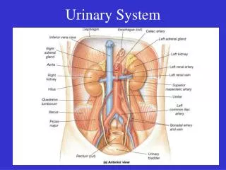
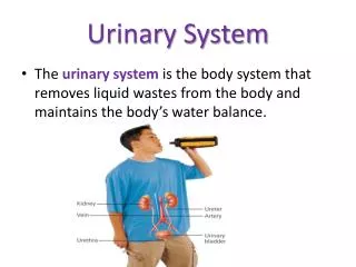
Urinary system By dr:Bakir.R.Rashid. Anatomy and functional evaluation of renal system. Anatomy : The renal system compose of two kidneys ,a ureter arise from each kidney ,and they end in urinary bladder from which urethra arise.

E N D
Urinary system By dr:Bakir.R.Rashid
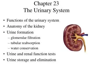
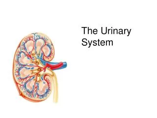
Anatomy and functional evaluation of renal system Anatomy : The renal system compose of two kidneys ,a ureter arise from each kidney ,and they end in urinary bladder from which urethra arise. The kidneys lie in the retroperitoneal space slightly above the level of the umbilicus.
-each kidney is about 6cm in length ,and 24gm in weight this is in full-term newborn , while in adult it is about 150gm and 12cm. -The functional unite of kidney is called nephron and each kidney contain approximately one million nephron.
Renal Function 1-Filtration, clearance and excretion of nitrogenous waste. 2-Fluid & electrolyte balance and acid-base hemostasis. 3-Endocrine organ elaborating many hormones like erythropoietin, prostaglandins, renin, vitamin D.
Approach to patient suspecting to have renal disorder History :renal disorder in pediatric age may be asymptomatic and discovered accedentally,or may present with nonspecific symptoms or signs like poor feeding, failure to thrive,irritability,poor weight gain, fever, diarrhea ,vomitnig,abdominal pain,abdomina mass, skin change or with color ,and odor change of urine. It may presents with anemia hypertension.
Physical examinations: Complete physical examination should be done with vital signs especially blood pressure with anthropometric measure.
Investigations Urinalysis (GUE) :although not totally specific or sensitive, is a useful, non invasive indicator of renal function and disease. Urine bags can be used to collect urine in small infants, however, they should never be used to obtain a culture. A-Urine concentration and dilution can be measured by specific gravity or osmolality. Normal urine specific gravity is 1003-1035.
B- Urine dipsticks provide a general estimate of: -acidity(normal PH 5-7). • albumin( up to 150mg/24hr). • glucose (normally no sugar in urine). • ketones (normally no ketone in urine). • bilirubin (indicate liver disease). • RBC (0-4). • pus cells which is a dead WBC normally there is (1-5 ) pus cells under high power field ,higher value indicate urinary tract infection. • nitrite in the urine indicate infection of urinary tract by gram negative bacteria.
C- Urine microscopy. A freshly voided urine specimen is centrifuged and examined for: 1. bacteria. It is difficult to distinguish infecting organisms from contaminants, also pyuria is not a reliable indicator of infection because infection may occur in the absence of pyuria, and pyuria may occur with acute illness in the absence of infection.
2. Cells:The morphology of RBCs in urine may help distinguish glomerular bleeding from blood loss elsewhere in the urinary tract. Crenated, dysmorphic RBCs in fresh urine suggest a glomerular origin.
3. Casts: of different types: (RBC casts) are the result of glomerular bleeding and are usually diagnostic of glomerulonephritis. ( Leukocyte casts): are occasionally seen in pyelonephritis and interstitial nephritis. (Hyaline casts and granular cats) :are not diagnostic of renal disease and may occur in the sediment of children who have oliguria of any cause.
4. Crystals:of many varieties may be present in urine they are rarely diagnostic of disease. An exception is the hexagonal cystine crystals, which is diagnostic of cystinuria.
Test of glomerular function: A. GFR: The creatinine clearance test is used to estimate the glomerular filtration rate (GFR). However, because a small amount of creatinine is released by the filtering tubes in the kidneys, creatinine clearance is not exactly the same as the GFR. In fact, creatinine clearance usually overestimates the GFR. This is particularly true in patients with advanced kidney disease. Clearance is often measured as milliliters/minute (ml/min). In young children and infants creatinine clearance can be estimated as follows: CC = K(constant)x(Ht. in cm.)/s.Craetinine in mg/dL. K=0.45 in infants <1year, 0.55 in children>1year, 0.7 in adolescent. Normal values are: •Male: 97 to 137 ml/min. •Female: 88 to 128 ml/min.
B. Urinary protein excretion: total protein excretion is <150mg/24hours protein to creatinine ratio should be < 0.2. A ratio exceeding 3.0 suggest heavy proteinuria of nephrotic syndrome.
Creatininein the urine: A random urine sample or a 24-hour collection may used. This test can be used as a screening test to evaluate kidney function. It may also be used as part of the creatinine clearance test. It is often used to provide information on other chemicals in the urine such as albumin or protein. Urine creatinine (24-hour sample) values can range from 500 to 2000 mg/day. Results depend greatly on your age and amount of lean body mass. Another way of expressing the normal range for these test results are: •14 to 26 mg per kg of body mass per day for men •11 to 20 mg per kg of body mass per day for women
Test of renal tubular function: A- Concentrating, diluting, and acidifying capacity. Renal concentrating capacity is assessed by a well monitored overnight fluid deprivation test, with serial determination of acute weight loss, serum osmolality, urine osmolality, and maximum urine specific gravity
- Urine concentration and dilution can be measured by specific gravity or osmolality. Normal urine specific gravity is 1003-1035.
-Urine osmolality is a measure of urine concentration, it is more accurate than specific gravity. -The 24-hour urine osmolality should be, on average, 500-800 mOsm/kg of water. After 12-14 hours of fluid intake restriction, the urine osmolality should exceed 850 mOsm/kg of water.
The following are associated with increased urine osmolality: •Dehydration •Syndrome of inappropriate antidiuretic hormone secretion (SIADH) •Adrenal insufficiency •Glycosuria •Hypernatremia •High-protein diet
Decreased urine osmolality is associated with the following: •Diabetes insipidus •Excessive fluid intake •Acute renal insufficiency •Glomerulonephritis
B- Tubular re absorptive dysfunction is suggested by detection of compounds in the urine that are normally reabsorbed completely by the renal tubules such substances include glucose, amino acids, and B2-microglobulin
BLOOD TESTS -blood uria nitrogen test (BUN): Urea nitrogen is what forms when protein breaks down. A test can be done to measure the amount of urea nitrogen in the blood. The normal result is generally 6 - 20 mg/dL. Lower-than-normal levels may be due to: •Liver failure •Low protein diet •Malnutrition •Over-hydration
- Serum Creatinine: Creatinine is a breakdown product of creatine, which is an important part of muscle. A normal result is 0.7 to 1.3 mg/dL for men and 0.6 to 1.1 mg/dLfor women. Lower than normal levels may be due to: -Muscular dystrophy (late stage) -Myasthenia gravis
Imaging procedures Ultrasonography: provides information on kidney location, size, shape and consistency. It can be used to diagnose obstruction, malformation, cysts, calcification, and tumors When combined with Color Doppler Imaging blood flow velocity in the renal artery and renal vein can be evaluated. Retrograde voiding cystourethrography: The contrast material is instilled by urethral catheter, and the bladder is visualized using fluoroscopy. It defines the presence and magnitude of vesicoureteral reflux and provides information about the capacity and anatomy of the bladder and urethra.
Radionuclide scanning :can provide an estimate of total renal function including GFR with the use of Technetium 99m labeled diethylenetriaminepenta-acetic acid, as well as tubular function with use of mercaptoacetyltriglycerine(MAG3), this can be used to provide cystograms in the assessment of vesicoureteral reflux. Technetium 99m labeled dimercaptosuccinic acid localizes to tubular cells of functioning nephrones, is very valuable in the diagnosis of renal parenchymal infection and scarring by demonstrating focal areas of decreased uptake.
Renal biopsy Is the definitive study for histological diagnosis of renal disease. It provides tissue for examination by light, immunofluorescence, and electron microscopy. Contraindications to percutaneous biopsy include bleeding disorders and the presence of a single kidney. The risk of the procedure include obtaining insufficient tissue for diagnosis, causing bleeding or infection, and creating an arteriovenous fistula within the kidney.