Download

1 / 24
240 likes | 444 Views
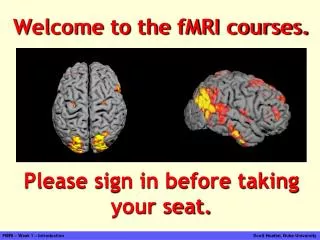
fMRI – difficulties in mind reading. fMRI: functional (nuclear) magnetic resonance imaging Neuroimaging: get the structure of the brain Want to know how it works: connection brain parts and brain functions Aim: measure the local „thinking activity” Usage (criticism): Lie detector

E N D
fMRI: functional (nuclear) magnetic resonance imaging • Neuroimaging: get the structure of the brain • Want to know how it works: connection brain parts and brain functions • Aim: measure the local „thinking activity” • Usage (criticism): • Lie detector • Neural- and psychological-modell checking(think/know experiment)
How we use it • Human attempts frequent • Well-planned tasks or questions • Measures: order of minutes • One measure: order of 5 seconds • Measure with and without tasks or question, further investigation based on the difference • Overlap the intensity map and brain image
The basics of fMRI • MRI: interaction between spins and magnetic field • QM based phenomena • Classical view is almost satisfactory • Find a „think-activity”-sensitive MRI measureable quantity, measure it, and then reflect to think activity
Get the signal - BOLD • Blood-oxygen-level dependence • Hemoglobin: Fe2+ can absorb O2 • Hemoglobin + O2 : diamagnetic molecule • Hemoglobin – O2 : paramagnetic (S=2) • Measure the oxygen-flow differencies in vein • Determine the connection between BOLD singal and toughts
Get the signal – O2 flow • The oxygen flow depends on the communicating intenstiy • Communication needs energy • Neuron cells don’t have repository • Increased activity needs more energy • BOLD signal decreasing and then increasing
Time, accuracy and resolution • Time: depends on the BOLD signal, about less than half a minute • Do experiments with the same patient, same time • Accuracy: easily detect maximum of BOLD • Space: resolution: in order of mm×mm×mm • Problem: the motion of the patient
Increasing space resolution • No new information with increasing resolution • Signal comes from multiple capillars • Solution: BOLD signal minimum: more localised • Longer time or • Bigger magnetic field • No (?) news: cerebral tasks are not well localised
Temporal resolution • Increasing time resolution does count • (more details) usable image • Two tricks to improve: • Spin echo – gradient echo • EPI: echo planar imaging
Measurement: EOM • First: apply • Short RF pulse:
Measurement: relaxation and decoherence • Solutionsafter„excitation”: • Wecanmeasure • Important: • New rotatingcylindricalcoordinateswithfrequency:
Measurement: slice-excitation and spin-echo • Find for the desired excitation-distribution:
Measurement: EPI • Echo Planar Imaging: increase time resolution even more!
An experiment: caffeine • caffeine as a contrast booster
Limits • We cannot answer any why question – only answer the question where • The brain is 3D – the image is 2D, so more experiments needed • We can measure only the neurons firing, but not the real actyvity ( block or stimulate?) • High degree of cerebral plasticity: places may vary
Mind reading • The map of opticnervestovissualcortex is so „localised”, „continous” • Trainingset: knownvideos and thosefMRIsignal • Unknown video: fMRIsignal → videoimages • Abilitytoguesswhatpatientthink • Numericalproblem • Furtheraim: movement of implants • Success: camera-eye