Download
1 / 9
100 likes | 463 Views
ABO and Rh Typing Procedure. PRINCIPLE AND APPLICATIONS .

E N D
PRINCIPLE AND APPLICATIONS • The ABO system is the most clinically significant blood group system for transfusion practice, because it is the only blood group system in which antibodies are consistently and predictably present in the serum of normal individuals whose red cells lack the antigen. ABO compatibility between donor and recipient is the foundation upon which all other pretransfusion testing rests. • The D(Rho) antigen is, after A and B, the most important red cell antigen in transfusion practice due to its potent antigenicity. Unlike the ABO system, however, individuals who lack the D antigen do NOT consistently and predictably have anti-D in their serum. • The ABO and Rh is determined for all patients who are candidates for transfusion, for all blood donors, for all prenatal patients, and for all potential organ recipients and donors.
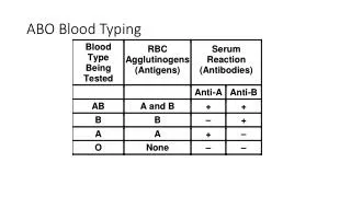
PROCEDURE • Verify that patient information on the sample matches information on the worksheet. • Centrifuge the sample and remove the serum to a labeled tube. • Add 2-3 drops of the patient’s cells to the WC tube, and prepare a washed 3% suspension. • Add 2 drops of patient serum to both the A1 and B tubes. This will be used for the serum confirmation, also known as the reverse grouping or backtyping.
Add one drop of reagent anti-A to the A tube, one drop of reagent anti-B to the B tube, one drop reagent anti-D to the D tube. • Add one drop of washed patient cells to: A tube, B tube, D tube. • Shake all tubes gently to mix. • Centrifuge tubes in the serofuge for the calibrated time
Gently resuspend each cell button individually and examine for hemolysis or agglutination with the aid of the lighted agglutination viewer. Grade results neg to 4+. • Immediately record the results in the appropriate spaces on the worksheet. • If the Rh test is negative, add a second drop of anti-D, then centrifuge and read again. • Discard all materials in the isolation trash containers.
WASHED 3% CELL SUSPENSIONS • PRINCIPLE • Washing cells to be tested removes serum or plasma which may contain proteins that interfere with testing, causing non-specific agglutination or rouleaux formation. Washing also removes fibrinogen, which may cause small clots. • The ratio of serum to cells markedly affects the sensitivity of agglutination tests. Preparation of a 2-5% cell suspension provides cells in an optimum concentration to detect weak antibodies.
SAMPLE • Washed 3% suspensions may be made from clotted whole blood, from anticoagulated patient samples, or from donor blood taken from a sealed segment on the donor unit.
Procedure • Using an indelible marking pen, label tube with the last name of the patient whose cells are to be washed • Using a dispopipet, transfer 2-4 drops of blood from the sample to the labeled tube. • Forcibly squirt saline from the wash bottle into the tube until it is about 3/4 full. • Centrifuge 45-60 seconds at high speed in the serofuge.
Decant into the dump bucket and shake to resuspend completely. • If gross hemolysis is present, repeat steps 3 to 5 until supernate is reasonably clear. • After the final wash, shake the tube to completely resuspend the cells and add saline to a final concentration of approximately 3%. Reagent test cells are in a 2-5% concentration. These can be used as a comparison.