Download
1 / 50
500 likes | 596 Views
UTERINE LEIOMYOMATA. Dr Zeinab Abotalib MD, MRCOG Associate Professor & Consultant Obstetrics & Gynecology Infertility And Assisted Conception. Uterine Leiomyomata. Benign tumor comprised mostly of smooth muscle cells First described by Reinier De Graff 1641

E N D
UTERINE LEIOMYOMATA Dr Zeinab Abotalib MD, MRCOG Associate Professor & Consultant Obstetrics & Gynecology Infertility And Assisted Conception
Uterine Leiomyomata • Benign tumor comprised mostly of smooth muscle cells • First described by Reinier De Graff 1641 • Most common tumor of the female pelvis • Represent 1/3 of all GYN admissions to hospitals
Incidence • Usually quoted 50% (Underestimate) • Cramer and Patel • 100 serial Uteri • Sectioned at 2mm • 77 of 100 had myomas • 84% had multiple myomas • 649 myomas found in all • No difference in incidence within pre or post menopausal uteri Am J Clin Pathol. 1990 Oct;94(4):435-8
Incidence • More common in African-Americans than white • Torpin et al. investigated 1741 Uteri • Overall incidence 3 times higher in blacks • Also tended to be larger • Also occurred at a younger age J Obstet Gynecol 1942;44:569
Incidence • Cumulative incidence by age 50, > 80% for African American and nearly 70% for Caucasian women. • One in four women have at least one submucosal fibroid. • Overall prevalence of uterine fibroids increases with age from 3.3% in women 25-32 to 7.8% in women 33-40 years. • Baird et al, Am J Obstet Gynecol 2003. • Borgfeldt et al, Acta Obstet Gynecol Scand 2000.
Etiology • Arise from a single muscle cell (monoclonal). • Proliferate under the influence of sex hormones, including estrogen, progesterone & androgens. • Effects of steroids are modulated by local growth factors. • Rein et al, Am J Obst Gyne 1995. • Ichimura et al, Fertil Steril 1998. • Stewart et al, Obstet Gynec 1998. • Wer et al, Fertil Steril 2002.
Etiology • Fibroblast growth factor • Vascular endothelial growth factor • Heparin-binding epidermal growth factor • Platelet-derived growth factor • Transforming growth factor • Parathyroid hormone-related protein • Prolactin
Etiology • Risk Factors • Nurses Health Study II • 95,061 nurses completed questionnaires in 1989, 1991, 1993 • Obesity • Early menarche • Nulliparity Fertil Steril. 1998 Sep;70(3):432-9
Etiology • Oral Contraceptives • High dose pills have been assoc. with stimulation of fibroid tumors • Smoking
Presentation • Most fibroids do not cause symptoms. • 20-50% experience tumor-related symptoms: • Menstrual dysfunction • Bowel and bladder dysfunction • Bulk effects • Such symptoms, account for up to 35% of all hysterectomies. • Lefebvre et al, J Obstet Gynecol Can 2003. • Myers et al, Agency for Health Care Research and Quality, 2001.
Pelvic Pain Menstrual Irregularities GI complaints Bladder complaints Dyspareunia Back pain Leg pain Vascular symptoms Infertility Asymptomatic Symptoms
Diagnosis • History • Bimanual pelvic or abdominal exam • Pelvic ultrasound - most common • MRI, HSG, sonohysterogram, hysteroscopy
Degenerative Changes • Degenerative changes are reported in approximately two-thirds of all specimens, but most of them have no clinical significance. • Hyaline degeneration- It is the most common • Cystic degeneration • Mucoid degeneration • Fatty degeneration • Carneous degeneration • Calcification • Sarcomatous degeneration(malignant transformation)
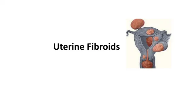
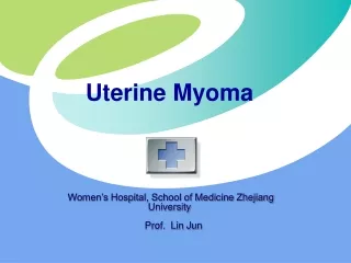
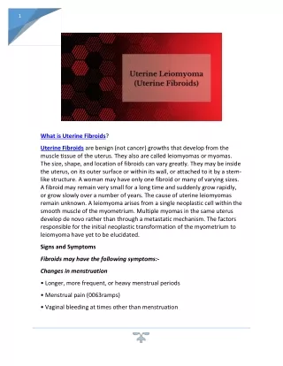
Uterine Fibroids Benign tumour of uterine tissue 3 locations: • subserosal • intramural • submucosal
Leiomyomas classified according to their location in the uterus
How are they diagnosed? • Usually detected on an internal gynecological exam • Diagnosis is usually confirmed by ultrasound but can also be made with magnetic resonance (MR) or computed tomography (CT) scans.
Factors that should be considered prior to initiating treatment include: • Size of the myoma(s) • Location of the myoma(s) (Symptoms • Woman's age (eg, is she near menopause?) • Reproductive plans
How are they treated? • Depends on size and location • Surgical therapy - hysterectomy, myomectomy • Drug therapy - pain relievers, hormone therapy (to shrink them) • Uterine artery embolization
Treatment • Expectant management - most cases • Indications for treatment • Abnormal uterine bleeding, causing anemia • Severe pelvic pain • Large or multiple • Obscuring evaluation of adnexa • Urinary tract symptoms • Postmenopausal or rapid growth
Treatment Choices • Medical therapies • Medroxyprogesterone (Provera) • Danazol • GnRH agonists (nafarelin acetate, Depot Lupron)
Treatment • RU486 • Anti-progestin • High affinity to Progesterone and glucocorticoid receptors • Murphy et al (1995) showed decrease of volume an average 49% • Recent reviews supports usage, but has been associated with • Hot flashes • Endometrial hyperplasia • Is not associated with trabecular bone loss Fertil Steril. 1995 Jul;64(1):187-90 Obstet Gynecol. 2004 Jun;103(6):1331-6 Clin Obstet Gynecol. 1996 Jun;39(2):451-60
Treatment • Gestrinone • Antiestrogen/antiprogesterone • GnRH analogues • Suppresses pituitary mediated secretion of estrogens • Basically treat 3-6 months • Expect 50% reduction of uterine volume
Treatment Choices • Uterine Artery Embolization (UAE)
UAE • Within three months following embolization: • 45% and 55% reduction in total uterine and myoma volume. • Reduction in symptoms in approximately 80% of women. • long- term data on durability and effects on fertility and pregnancy outcomes are very limited. Pron et al, Fertil Steril 2003 Burbank et al, J Am Assoc Gynecol Laparosc 2000
Myomectomy • First performed by Washington and John Atlee, 1844 • May be approached in a variety of ways • Abdominally (open) • Laparoscopic • Hysteroscopic • Primarily for submucosal/intramural fibroids impacting the endometrial cavity • Vaginal • Primarily for pedunculated submucous fibroids
Myomectomy • Biggest complication is blood loss
Myomectomy (local surgical removal of fibroids) • Sparing the uterus • Complications significant blood loss could require hysterectomy • Fibroids can recur20 - 25% will need another procedure for treatment of new fibroids
Myomectomy • Hysteroscopy for intracavitary / submucous • Laparotomy
Myomectomy • Hysteroscopy for intracavitary / submucous • Laparotomy
Treatment Choices • Hysterectomy • Vaginal • Abdominal
Hysterectomy • Curative, but irreversible • Until now, the standard therapy for fibroids 1/3 of all hysterectomies are performed for fibroids • Complications: bleeding, infection, adhesions, risks associated with general anesthetic • 6 - 8 week recovery
Method Of Delivery Vertex- Vertex (50%) • Vaginal delivery, interval between twins not to exceed 20 minutes. Vertex- Breech (20%) Vaginal delivery by senior obstetrician
Method Of Delivery Breech- Vertex( 20%) • Safer to deliver by CS Breech-Breech( 10%) • Usually by CS.
Method Of Delivery • MONO-MONO • By C/S • Why?