Download
1 / 30
310 likes | 505 Views
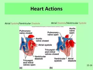
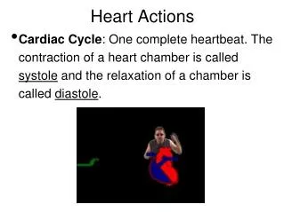
Heart Actions. Cardiac Cycle : One complete heartbeat. The contraction of a heart chamber is called systole and the relaxation of a chamber is called diastole . . 1 complete sequence of pumping heart contracts & pumps heart relaxes & chambers fill contraction phase systole

E N D
Heart Actions • Cardiac Cycle: One complete heartbeat. The contraction of a heart chamber is called systole and the relaxation of a chamber is called diastole.
1 complete sequence of pumping heart contracts & pumps heart relaxes & chambers fill contraction phase systole ventricles pumps blood out relaxation phase diastole atria refill with blood Cardiac cycle
Stethoscope - instrument to listen and measure heart sounds Heart Sounds - Opening and Closing of Valves, "Lub Dub"
Lub-dub, lub-dub • Heart sounds- closing of valves • “Lub” • force blood against closed AV valves • signifies beginning of systole • “Dub” • force of blood against semilunar valves • close at the beginning of ventricular diastole SL AV AV
Heart murmur • Irregular heart sounds • leaking valve causes hissing sound • blood squirts backward through valve
The cusps (flaps) of the bicuspid and tricuspid valves are anchored to the ventricle walls by fibrous “cords” called chordae tendineae, which attach to the wall by papillary muscles. This prevents the valves from being pushed up into the atria during ventricular systole. Can you identify these parts?
7. Papillary Muscle8.Chordae Tendinae9. Mitral Valve cusps • Right Atrium • Right Atrioventricular Valve (Tricuspid Valve) • Right Ventricle • Left Atrium • Left Atrioventricular Valve (Mitral Valve) • Left Ventricle
Blood pressure is the force of blood against the walls of arteries. Blood pressure is recorded as two numbers—the systolic pressure (as the heart beats) over the diastolic pressure (as the heart relaxes between beats).
How is this reflected in blood pressure measurements? ventriclesfill systolic ________ diastolic chambers fill pump(peak pressure) _________________ fill(minimum pressure) 110 ________ 80 ventriclespump
The average (normal) blood pressure for an adult is 120/80. This number varies by person and it is best if you know what is *normal* for you, so that you (or your doctor) recognize when something is not normal. We will be doing a lab where you will learn to use a this device and check your own blood pressure. SPHYGMOMANOMETER
Measurement of blood pressure if systolic > 150 or if diastolic > 90 hypertension =(high blood pressure)
Factors affecting blood pressure: Average is 120/80 (higher number is the systolic pressure) 1. Cardiac Output 2. Blood volume (5 liters for avg adult) 3. Blood Viscosity 4. Peripheral Resistance
Cardiac Output (CO) • Volume of blood pumped by each ventricle in one minute • CO = heart rate (HR) x stroke volume (SV) • HR = number of beats per minute • SV = volume of blood pumped out by a ventricle with each beat
Cardiac Output (CO) • At rest • CO (ml/min) = HR (75 beats/min) SV (70 ml/beat) = 5.25 L/min • Maximal CO is 4–5 times resting CO in nonathletic people • Maximal CO may reach 35 L/min in trained athletes • Cardiac reserve: difference between resting and maximal CO
Electrical signals • heart pumping controlled by electrical impulses • signal also transmitted to skin = EKG stimulates ventricles to contract from bottom to top, driving blood into arteries allows atria to empty completely before ventricles contract
Cardiac Conduction S-A Node Junctional Fibers A-V Node A-V Bundle Perkinje Fibers
1 Sinoatrial node (Pacemaker)2 Atrioventricular node3 Atrioventricular Bundle (Bundle of His)4 Left & Right Bundle branches5 Bundle Branches (Purkinje Fibers)
View the heart animations at McGraw Hillto understand the Cardiac Cycle
Regulation of Cardiac Cycle controlled by the cardiac center within the medulla oblongata. The cardiac center signals heart to increase or decrease its rate according to many factors that the brain constantly monitors. Muscle Activity Body Temperature Blood ion levels (potassium & calcium)
Variables in Heart Rate Increased muscle activity = Higher oxygen demands = increase in heart rate Higher Body temperature = increased heart rate; Lower body temp = lower heart rate Blood level of certain ions - Potassium High = Lower heart rate; Potassium Low = Higher heart rate - Calcium high = Higher heart rate; Calcium Low = Lower heart rate
ECG – electrocardiogram – a recording of the electrical events (changes) during a cardiac cycle • P Wave – depolarization of the atria (atrial contraction – systole) • QRS Complex – depolarization of the ventricles (ventricular contraction, systole) • T Wave – Repolarization of the ventricles Heart Sounds – opening and closing of the valves, flow of blood into and out of the chambers, vibrations in muscle
Irregular Heart Rates Arrhythmia-irregular heart rate Tachycardia- rapid heartbeat (>100 BPM) Bradycardia-slow heartbeat ( < 60 BPM) Fibrillation = rapid, uncoordinated unsynchronized heart rate. Atria (not serious. Ventricles (deadly)
SADS = (Sudden Arrhythmia Death Syndromes or Sudden Adult Death Syndrome) Routine ECG Screening may help prevent deaths in young people
Interpreting ECGs An ECG is printed on paper covered with a grid of squares. Notice that five small squares on the paper form a larger square. The width of a single small square on ECG paper represents 0.04 seconds. A common length of an ECG printout is 6 seconds; this is known as a "six second strip."
Analyze an ECG Each one of the figures represents an ECG pattern displaying three types of abnormal rhythms. Identify: Arrhythmia Tachycardia Bradycardia
Defibrillator common treatment for life-threatening cardiac arrhythmia The device shocks the heart and allows it to re-establish its normal rhythm The device can also be used to start a heart that has stopped.