Download
1 / 90
1.39k likes | 3.79k Views
Venous Access. Matthew L. Paden, MD Emory University Children’s Healthcare of Atlanta at Egleston. Peripheral IV. Butterfly & angiocaths Short catheters generally placed in forearm, hand or scalp veins Short term therapy and unable to handle caustic chemicals (chemotherapy).

E N D
Venous Access Matthew L. Paden, MD Emory University Children’s Healthcare of Atlanta at Egleston
Peripheral IV • Butterfly & angiocaths • Short catheters generally placed in forearm, hand or scalp veins • Short term therapy and unable to handle caustic chemicals (chemotherapy)
Peripheral Sites • Veins of the Forearm • 1. Cephalic vein2. Median Cubital vein3. Accessory Cephalic vein4. Basilic vein5. Cephalic vein6. Median antebrachial vein
Peripheral Sites • Veins of the Hand • 1. Digital Dorsal veins2. Dorsal Metacarpal veins3. Dorsal venous network4. Cephalic vein5. Basilic vein
Peripheral IVs • Try to cannulate the most distal veins first • Drugs or fluids put through the cannula may extravasate at the upstream failed cannula site • Transillumination • Topical nitropaste
Types of Central Vascular Access Devices • Non-tunneling • Tunneling • Implanted
Non-Tunneling • Direct venipuncture through the skin into a selected vein. • Peripheral IV • Peripherally inserted central catheter • Percutaneous catheters
Non-Tunneling - PICC • Peripherally inserted central catheters (PICC) • Midline • Central venous catheter inserted at or above the antecubital space and then advanced until the distal tip of the catheter is positioned at the superior vena cava or superior vena cava and right atrial junction.
Useful for patient receiving long term medication therapy, chemotherapy or TPN Used for frequent blood sampling Distal end positioned at the superior vena cava or junction of superior vena cava and right atrium Non-tunneling - PICC
Non-Tunneling - PICC • Peripherally inserted central catheters (PICC)
Non-Tunneling - Midlines • Used for shorter term intravenous therapy (up to 4 weeks) • Used for frequent blood sampling • Distal end positioned at the proximal end of the upper extremity
Non-Tunneling – PICC and Midline examples at the antecubital & above
Non-Tunneling – CVC • Percutaneous catheters • Also known as: Central Venous Catheters (CVC) • Subclavian, femoral or internal jugular • Single, double or triple lumen
Tip advanced to superior vena cava or SVC and right atrium junction As with PICC, appropriate for patients requiring long term chemotherapy or TPN Non-tunneling - CVC
Tunneling • Hickman® • Broviac® • Groshong®
Tunneling • Inserted into a central vein via percutaneous venipuncture or cut down • Catheter then tunneled under the skin in the subcutaneous tissue and exited in a convenient location • Dacron cuff hold the catheter in place
Tunneling - Broviac® • Similar to the Hickman catheter, but is of smaller size. • This catheter is mostly used for pediatric patients.
Tunneling - Groshong® • Similar to Hickman®and Broviac®with closed ended patented 3-way valve.
Implanted VADs - Ports • Catheter attached to a self-sealing silicone septum surrounded by a titanium, stainless steal or plastic port • Port sutured under the skin • Some brand names: • Port-a-cath® • Infus-a-port® • Power Port ®
Implanted VADs - Ports • Catheter runs from port to superior vena cava at the right atrium • No part of the device is exposed outside the body • Can deliver chemotherapy, TPN, antibiotics, blood products and blood sampling
Can only be accessed with special needle called a HUBER needle Contains a deflecting, non-coring point Implanted VADs - Ports
Insertion Complications • Inadvertent Arterial Puncture • Hematoma Formation • Extravasation • Infection • Phlebitis • Pneumothorax
Systemic Complications • Infection • Deep Vein Thrombosis • Pulmonary Embolism • Superior Vena Cava Syndrome
Mechanical Complication • Catheter tip migration • Broken or damaged catheter • Catheter occlusion
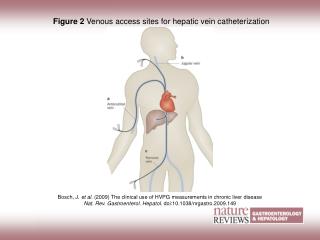
Femoral Vein • Adults – • DVT • Excess infection risk • “Potentially inaccurate CVP”
Femoral Vein • Kids – • Better risk profile • Ease of insertion, compressible • No difference in DVT – ref 1-2 • Same infection risk (maybe lower) – ref 3-5 • Accurately reflects RAP if no increase in abdominal pressures – ref 6-8 1. Beck C, et al. J Ped 1998;133:237-41. 2. Jacobs B, et al. Crit Care Med 1999;27:A29 3. Casado-Flores J, et al. Ped Crit Care Med 2001;2:57-62. 4. Richards M, et al. NNIS Pediatrics 1999;103:103-9 5. Stenzel JP, et al. J Ped 1989;114:411-5. 6. Fernendez E, et al. Ped Crit Care Med 2004;5:14-18 7. Lloyd R, et al. Pediatrics 1992;89:506-8. 8. Ho K, et al. Crit Care Med 1998;26:461-4.
Femoral anatomy • Vein is medial to the artery • Froehlich’s theorem • Superficial distal to inguinal ligament, then dives deep • 0.5-2cm inferior to the inguinal ligament
Quiz Question • What are the anatomic landmarks to determine where to stick for the femoral vein in a pulseless patient? • A. 1/3 of the distance from the anterior superior iliac spine to the pubic tubercle • B. ½ the distance between the pubic tubercle and the anterior superior iliac spine • C. 1/3 of the distance from the pubic tubercle and the anterior superior iliac spine • D. None of the above
Quiz answer • D. None of the above • The femoral ARTERY lies ½ the distance between the pubic tubercle and the anterior superior iliac spine. • The femoral vein is 0.5-1.5 cm medial to this depending on the size of the patient.
Straight vs. Frog leg • “The optimal positioning of the leg can vary according to the preference of the operator.” • Discuss
Procedure • 30-45 degree angle to skin • 2 methods • Stick with negative pressure on syringe while entering and exiting • Insert needle, and only negative pressure on removal • Allows you to better stabilize the needle by resting your hand on the thigh
Procedure • Blood flash - Insert wire • Wire not going smoothly • Needle no longer in vessel • False tracking in subcutaneous tissue • Thrombus • Advancing into lumbar veins • Small incision • Blade directed away from wire
Procedure • Twisting motion of dilation • Remove dilator • Advance catheter • Remove wire • Aspirate and flush all ports • Secure line with sutures • Sterile dressing
Procedure • Wheeler – “Confirmation of proper CVC position is required after placement of all CVC’s”
Warnings • If you hit the artery – pressure until hemostatic • Wire should float – should never have resistance • If can’t pull the wire through the needle – remove both wire and needle together so you don’t sheer off the wire • Never let go of the wire • Catheter tip “pointing too cephalad” – in lumbar veins
Complications • 74 of 89 (83%) – no complications • Other 15 – minor bleeding/hematoma • 94.4% success rate • Median duration 5 days • 21% <3 days 26% 7-14 days • 43% 4-7 days 10% >14 days • Long term – 8 leg swelling, 11 BSI Venkataraman, et al. Clin Ped 1997;36:311-9.
Complications • 45 months – 395 CVL – 162 femoral • No insertion complications • Mean duration 8.9 days • 9 noninfectious complications • 4 thrombosis, 1 perforation, 1 embolism, 2 bleeding • “The low incidence of complications in this study suggests that the femoral vein is the preferred site in most critically ill children when CVC is indicated.” Stenzel JP, et al. J Ped 1989;114:411-5