Download
1 / 27
290 likes | 315 Views
Second trimester miscarriage and cervical insufficiency. Dr. HIND A. SHOWMAN. Definition.

E N D
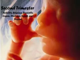
Second trimester miscarriage and cervical insufficiency Dr. HIND A. SHOWMAN
Definition • Spontaneous miscarriage is defined as the spontaneous loss of a pregnancy prior to viability, taken as 23 weeks and 6 days of gestation. Beyond this gestation, fetal demise is classified as a stillbirth. The majority of first‐trimester miscarriages occur below 12 weeks’ gestation with an overall rate of around 20%. Second‐ trimester miscarriages are less common, accounting for 1–4% of all miscarriages.
Aetiology: • Although the causes of miscarriage in the first and second trimester appear different, there is inevitably some overlap • The likely Aetiology behind second trimester losses varies with gestational age. •At 12 -16 weeks, the predominant causes are those of first trimester miscarriage - fetal chromosomal and structural anomalies and possibly endocrine causes. •At the latter part of the second trimester, between 17 and 23 weeks, the commonest factors underlying the miscarriages will be those of very pre term births, these include ascending genital tract infection, intrauterine bleeding and cervical weakness. The weakness may be congenital or acquired through procedures such as cone biopsy or repeated cervical dilatation.
Uterine abnormalities: sub mucous fibroids and congenital distortion of the cavity (uterine septa) may be implicated. • Thrombophilia. • Epidemiological risk factors for second trimester miscarriage include poor socioeconomic status, smoking, genital tract infections, previous miscarriage or premature birth and increasing maternal age .Amniocentesis a procedure performed at 16 -18 weeks gestation is associate with o.5 % of subsequent pregnancy loss.
Investigations: • 1. CBC for anemia, leukocytosis, clotting screen. • 2. Blood consider Group Rh if –ve give anti D. • 3. Infection screen (HVS, endocervical swabs, MSU, CRP, blood cultures) should be performed if • maternal infection is suspected, particularly in the presence of pyrexia, flu-like symptoms, • abnormal liquor or prolonged rupture of membranes. • 4. Trans abdominal ultrasound to confirm gestation and viability, and assess amniotic fluid volume. • 5. Transvaginal ultrasound for cervical length measurement
Differential diagnosis: • 1-Bowel disorders: constipation , colitis ,gastroenteritis . • 2-Urinary disorders: infection ,renal stone disease. • 3-cervical disorders: ectropion ,leucorrhoea,Neoplasia . • 4- Uterine disorders: red degeneration of fibroids , round ligaments stretching .
Management • Management options fall into three groups, medical, surgical or expectant. Factors to be taken into account when discussing these options with patients include the following, type of miscarriage, gestation at which miscarriage is diagnosed. • • Support this include sympathy, explanations, pain relieve and reassurance. • • There is currently no place for tocolysis in the prevention of mid trimester pregnancy loss , tocolytics only used for short term i.e. 48 hours prolongation of pregnancy . • • Emergency cervical circulage when the cervical canal is opened and the membrane is bulging it is suitable to insert suture around the cervix to close it after reducing the membrane .Bleeding, infection and contractions are contraindications.
• If the pre viable membrane rupture precise diagnosis should be confirmed by speculum examination and follow up ultrasound scan if no contraction and no sign of infection no urgency to interfere but severe oligohydramnia carry risk of fetal lung hypoplasia .Carful observation of body temperature ,and changing vaginal discharge or lower uterine tenderness are recommended Established chorioamnitis needs delivery . • • Antibiotics should be given during follow up of the patient during expectant management • • Delivery : adequate pain relief should offered , these deliveries are usually vaginal and quick .
Cervical Insufficiency: • The cervix is a cylinder-shaped neck of tissue that connects the vagina and uterus. Located at the lowermost portion of the uterus, the cervix is composed primarily of fibromuscular tissue. There are two main portions of the cervix: • The part of the cervix that can be seen from inside the vagina during a gynecologic examination is known as the ectocervix. An opening in the center of the ectocervix, known as the external os, opens to allow passage between the uterus and vagina. • The endocervix, or endocervical canal, is a tunnel through the cervix, from the external os into the uterus. • The overlapping border between the endocervix and ectocervix is called the transformation zone. • The cervix produces cervical mucus that changes in consistency during the menstrual cycle to prevent or promote pregnancy. • During childbirth, the cervix dilates widely to allow the baby to pass through. During menstruation, the cervix opens a small amount to permit passage of menstrual flow.
Definition: • (formerly called cervical incompetence) is painless cervical dilation resulting in delivery of a live fetus during the 2nd trimester.
Risk Factors of cervical incompetenc • 1-Recurrent 2nd trimester losses • 2-History of incompetent cervix with a previous pregnancy • 3-Cervical injury -multiple D&C • -Repeated surgical trauma • - repeated pregnancy termination, • - cone biopsy • - cervical cautery (to remove growths or stop bleeding • 4-Anatomic abnormalities of the cervix • -congenital cervical hypoplasia or aplasia • 5-DES (diethylstilbestrol) exposure • 6-Connective tissue disorders (Ehlers-Danlossyndrome)
Incidence • Cervical incompetence affects 1 % of the obstetric population.
Diagnosis: • During pregnancy • -Based on an obstetric history of recurrent second- or early third-trimester fetal loss with the above criteria mentioned (painless cervical dilation). • -Sudden unexpected rupture of membrane followed by expulsion of the fetus • -Pelvic exam, examine cervix to see if the amniotic sac has begun to protrude through the opening (prolapsed fetal membranes). • - In addition to history, use assessment of cervical length in second trimester to identify cervical shortening using transvaginal ultrasound .However, short cervical length has actually been shown to be a marker of preterm birth rather than cervical incompetence. • Normally, the cervix should be at least 30 mm in length. Cervical incompetence is variably defined. However, a common definition is a cervical length of less than 25 mm at or before 24 weeks of gestational age
-- A normal sagittal view of the cervix shows a “T” shaped endocervical canal vs. deviations such as Y, V, U. Y= initial effacement and subsequent V, U visualized on progressive endocervial change and cervical shortening. TVS assessments in low-risk women to screen for cervical incompetance should not be done routinely. Management should be determined by prior history.
Or positive pre pregnancy physical finding: • 1-ability to introduce No 8 Hegar dilator through internal cervix when the patient not pregnant for assessment of the patulous cervix . • 2- hysterosalpingography demonstrating funneling of the cervix .
Indications for elective cerclage: • 1-congenital or acquired visible defects in the ectocervix • 2-classic features of cervical incompetence ,history of 2 or more 2nd trimester losses (excluding those resulting from preterm labor or abruption) • 3- history of losing each pregnancy at an earlier gestational age • 4- history of painless cervical dilation of up to 4 to 6 cm • 5-history of cervical trauma caused by cone biopsy, intrapartum cervical lacerations • 6- history of excessive, forced cervical dilation during pregnancy termination.
Contraindications of cervical cerclage: • 1. Uterine bleeding, • 2. ruptured membranes • 3. uterine contractions • 4. fetal anomaly • 5. chorioamnionitis • Timing of cerclage placement :at 13-16 weeks GA after fetal viability established on ultrasound • Timing of cerclage removal: should be an appropriate time before labor. at 37 weeks, if membrane ruptured ,labour started
Types of cervical cerclage • Macdonald cerclage procedure ;Running suture placed in the body of the cervix near the internal os to encircle the cervix . Its tightened to reduce the cervical canal to 5-10mm • Modified Shirodkar procedure More complicated and involving an anterior incision, placement and tying of special Mersiline tape with suturing of the cervical mucosa back in place. • reserved for patients that have had failure with the Macdonald procedure .
Complications: • 1.Suture disruption, • 2.rupture of membranes, • 3.chorioamnionitis