Download
1 / 23
250 likes | 485 Views
Dosimetry part 1: X-ray interactions with matter. G.Haddadi, PhD, MSc Associated prof. of Medical Physics Fassa University of Medical Sciences. Basic concepts of risk versus benefit. Radiation protection can only be practiced well when understood the risks and benefits of radiation

E N D
Dosimetrypart 1: X-ray interactions with matter G.Haddadi, PhD, MSc Associated prof. of Medical Physics Fassa University of Medical Sciences
Basic concepts of risk versus benefit Radiation protection can only be practiced well when understood the risks and benefits of radiation Minimizing the exposure while maximizing the diagnostic and therapeutic efficacy of an examination
پرتو گیری از منابع طبیعی و مصنوعی • رادن 55% • کیهانی 8% • رادیونوکلئیدهای زمینی 8% • رادیونوکلئیدهای داخل بدن 11% • پزشکی 15% • سایر موارد 3%
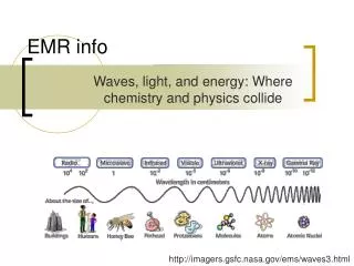
Interaction between X-rays and Matter • X-rays can interact with orbital electrons and nuclei of the atoms • In diagnostic energy range most interactions occur at orbital electron levels • Five ways of interactions: 1.Coherent scattering 2.Photoelectric effect 3.Compton scattering 4.Pair production, and 5.Photodisintegration
1. Coherent (Rayleigh) Scattering • Photon interaction with loosely bound-orbital electron • Low energy photons (hν << m0c2) • Photon elastic scattering • Incident Photon is deflected into another direction losing little energy • Negligible change in wave length • Negligible energy deposition • Sometimes reduces the resolution of image
2. Photoelectric Absorption • Interaction between lower energy photons and inner shell electrons (e.g “K” shell) • The total energy of an x or γ ray is transferred to an inner electron. • The electron is ejected from the atom with kinetic energy, Eκ; Eκ= hν − EB
Photoelectric effect yields • Ejected photoelectron • Characteristic radiation with energy of; hv=ΔE= E1 –E2 • Auger electrons with kinetic energy of; EK= hv-EB Electrons are ejected approximately at a right angle as low-energy photons interact photoelectrically. As the energy of the photons increases, the angle decreases between incident photons and ejected electrons.
Probability of photoelectric interaction and/or attenuation coefficient(ζ) • Decreases rapidly as the photon energy increases: ζ~ 1/(hν)3 • The photoelectric mass attenuation coefficient also depends to atomic number, it varies with Z3 ζ~ Z3 / (hν)3 Photoelectric mass attenuation coefficients of lead and soft tissue as a function of photon energy. K and L-absorption edges are depicted for lead.
X and γ rays with energy between 30 keV and 30 MeV interact in soft tissue predominantly by Compton scattering A Part of the energy of an incident photon is transferred Loosely bound or “free” electron 3. Compton Scattering (Incoherent)
Compton interaction yields • Compton electron; with deflection angle of θ; 90< θ<0 • Scattered photon; with scattering angle of φ; 180<φ<0 • change in wavelength, Δλ; Δλ = 0.00243 (1 − cosφ) Δλ = λ’- λ
Compton attenuation coefficient (σ) • The coefficient decreasesgraduallywith increasing photon energy • The Compton mass attenuation coefficient varies directly with the electron density (electrons per kilogram) • Almost, independent of the atomic number
Radiographic contrast comparisonfor different photon energies Radiographs taken at 70 kVp, 250 kVp, and 1.25 MeV (60Co). These films illustrate the loss of radiographic contrast as the energy of the incident photons increases.
4. Pair production:conversion of energy to mass • The nucleus is involved • A pair of electrons, one negative and one positive, appears in place of the photon • Mass of an electron and/or positron is 0.51 MeV • Energy threshold; hνmin= 1.02 (MeV)
Pair production cont’d • During pair production, energy in excess of 1.02 MeV is released as kinetic energy of the two electrons: hν (MeV) = 1.02 + (Ek)e− + (Ek)e+ • Annihilation photons are produced when the positron and an electron annihilate each other.
Pair production attenuation coefficient (k) • k varies almost linearly with the atomic number of the attenuating medium • Coefficient increases slowly with energy of the incident photons
Relative importance of the three principal interactions of x and γ rays
5. Photodisintegration • Complete absorption of incident high energy photon • Ejection of nuclear particles or nucleons such as neutron or protons
ATTENUATION OF X AND γRADIATION • Three possible outcomes upon impinges of x or γ ray: (1) Absorption (i.e., transfer its energy to atoms of the target material) during one or more interactions; (2) Scattering during one or more interactions; or (3) Traverse the material without interaction.
Photon attenuation dependes on: • The number of primary photons transversing the medium (I0) • Thickness of material (x) • Total attenuation coefficient of medium (μ) • For monoenergetic photons and narrow beam condition: I = I0e−μx
Total attenuation coefficient of medium (μ):μ = ω + τ + σ + κ + π Mass attenuation coefficients (in cm2/g), is obtained by dividing linear attenuation coefficients by the density ρ of the attenuating medium: μm=μ/ρ , τm=τ/ρ, σm=σ/ρ , κm=κ/ρ Mass attenuation coefficients for selected materials as a function of photon energy.
Attenuation and Half Value Layer (HVL) • The thickness of a slab of matter required to reduce the intensity (or exposure rate) of an x- or γ -ray beam to half • HVL describes the “quality” or penetrating ability HVL = ln 2/μ I = I0e−μx
The absorption coefficient is determined in terms of the Half-Value Layer HVLwhich is the thickness of a material necessary to reduce the intensity to 50% of its original value. The solution yields:
Graph showing how the intensity of an x-ray beam is reduced by an absorber whose linear absorption coefficient is = 0.10 cm1.