Download
1 / 18
210 likes | 558 Views
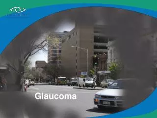
Glaucoma. Visual Field Examinations Week 5. Glaucoma. Glaucoma is a disease of the optic nerve. Progressive loss of axon or nerve fibers cause irreversible visual field loss.

E N D
Glaucoma Visual Field Examinations Week 5
Glaucoma • Glaucoma is a disease of the optic nerve. • Progressive loss of axon or nerve fibers cause irreversible visual field loss. • The area of visual field loss is determined by where the damage has occurred either on the rim of the optic nerve or the nerve fiber layer.
High tension glaucoma Pressure readings higher than the normal, 10-21mm hg will be observed. The higher than normal pressure readings will cause a loss of axons throughout the nerve with concentric enlarging of the cup and thinning of the rim. This type of damage causes a generalized decrease in sensitivity. This type of VF loss is seen in the early stages of glaucoma.
High tension glaucoma When viewing a VF automated printout, readings for the average “normal” VF should be in the lowers 30’s centrally and upper 20’s-lowers 30’s in the peripheral VF. A patient with high tension glaucoma will have lower than “normal” readings across the whole VF. Progressively, the high tension patient will have a “ring around the collar” effect. This "collar” will get progressively worse over the years and close in to tunnel vision and end stage glaucoma.
High tension glaucoma This is an example of high tension glaucoma shown over a period of years. A computer printout from the Humphrey VF allows the technician to printout a comparison of all VF’s done a one patient. This allows the doctor to see all of the VF’s done on a patient so he doesn’t have to flip through the chart to get the comparison.
Low tension glaucoma Patient’s with low tension glaucoma will have pressure readings within the “normal” limits of mm hg.( between 10-21mm Hg) The damage from low tension glaucoma may occur in a more focal manner, with enlargement of the cup toward the superior and/or inferior poles of the disc. This will result in nerve fiber bundle defects in the VF.
Low tension glaucoma Important note Red=macula Pink=defect • Nerve fiber bundle defects will always show opposite of the damaged area on the visual field. • If the defect is on the inferior retina, it will show superior on the VF.
Nerve fiber bundle defects Arcuate Arcuate
Arcuate nerve fiber bundle defect This nerve fiber layer has damage inferiorly but shows superiorly on the VF. This “reversal” will always happen with any defect before the chiasm. Any defect in the retina, macula, and nerve will show opposite of the damage on the VF.
Nerve fiber bundle defects Nasal step Nasal step
Nerve fiber bundle defects Temporal wedge Temporal wedge
Advanced glaucoma After years of glaucoma, the individual defects get worse. New nerve fiber bundle defects can occur and worsen. In advanced glaucoma or end stage glaucoma all of the individual nerve fiber bundle defects start to combine into one large defect, resulting in tunnel vision.