Download
1 / 59
590 likes | 653 Views
Solving protein structures,m olecular mechanics , and d ocking Lecture 18 Introduction to Bioinformatics 2006. Thursday May 4th. NO LECTURE But … 13:30 – 15:15 hrs in S329 and S345 : PRACTICAL HOMOLOGY SEARCHING. Today’s lecture.

E N D
Solving protein structures,molecular mechanics, and dockingLecture 18Introduction to Bioinformatics2006
Thursday May 4th NO LECTURE But … 13:30 – 15:15 hrs in S329 and S345: PRACTICAL HOMOLOGY SEARCHING
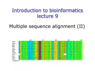
Today’s lecture • Experimental techniques for determining protein tertiary structure • Molecular motion simulated by molecular mechanics • Protein interaction and docking • Ribosome example • Zdock method
If you throw up a stone, it is Physics. If it lands on your head,it is Biophysics.
If you throw up a stone, it is Physics. If it lands on your head,it is Biophysics. If you write a computer program, it is Informatics.
If you throw up a stone, it is Physics. If it lands on your head,it is Biophysics. If you write a computer program, it is Informatics. If there is a bug in it, it is Bioinformatics
Experimentally solving protein structures Two basic techniques: • X-ray crystallography • Nuclear Magnetic Resonance (NMR) tchniques
Phase problem Crystallization 1. X-ray crystallography Purified protein Crystal X-ray Diffraction Electron density 3D structure Biological interpretation
Protein crystals • Regular arrays of protein molecules • ‘Wet’: 20-80% solvent • Few crystal contacts • Protein crystals contain active protein • Enzyme turnover • Ligand binding Example of crystal packing
Examples of crystal packing Acetylcholinesterase ~68% solvent 2 Glycoprotein I ~90% solvent (extremely high!)
hydrophilic Lipid bilayer Flexible hydrophobic hydrophilic Flexible and heterogeneous!! Problematic proteins (no crystallisation) • Multiple domains • Similarly, floppy ends may hamper crystallization: change construct • Membrane proteins • Glycoproteins
Liq.N2 gas stream X-ray source beam stop detector goniometer Experimental set-up • Options for wavelength: • monochromatic, polychromatic • variable wavelength
Diffuse scattering (from the fibre loop) Water ring Direct beam Beam stop Reflections (h,k,l) with I(h,k,l) Increasing resolution Diffraction image reciprocal lattice (this case hexagonal)
The rules for diffraction: Bragg’s law • Scattered X-rays reinforce each other only when Bragg’s law holds: Bragg’s law: 2dhkl sin q = nl
Phase Problem • Determining the structure of a molecule in a crystalline sample requires knowing both the amplitude and the phase of the photon wave being diffracted from the sample • X-rays which are emitted start out with dispersed phases, and so the phases get lost • Unfortunately, phases contribute more to the informational content of a X-ray diffraction pattern than do amplitudes. It is common to refer to phaseless X-ray data as having "lost phases“ • Luckily, several ways to recover the lost phases have been developed
Building a protein model • Find structural elements: • -helices, -strands • Fit amino-acid sequence
Building a protein model • Find structural elements: • -helices, -strands • Fit amino-acid sequence
d = 4 Å Effects of resolution on electron density Note: map calculated with perfect phases
d = 3 Å Effects of resolution on electron density Note: map calculated with perfect phases
d = 2 Å Effects of resolution on electron density Note: map calculated with perfect phases
d = 1 Å Effects of resolution on electron density Note: map calculated with perfect phases
Refinement process • Bad phasespoor electron density maperrors in the protein model • Interpretation of the electron density map improved modelimproved phasesimproved map even better model … iterative process of refinement
Validation • Free R-factor (cross validation) • Number of parameters/ observations • Ramachandran plot • Chemically likely (WhatCheck) • Hydrophobic inside, hydrophilic outside • Binding sites of ligands, metals, ions • Hydrogen-bonds satisfied • Chemistry in order • Final B-factor (temperature) values
2. Nuclear Magnetic Resonance (NMR) 800 MHz NMR spectrometer
Nuclear Magnetic Resonance (NMR) • Pioneered by Richard R. Ernst, who won a Nobel Prize in chemistry in 1991, FT-NMR works by irradiating the sample, held in a static external magnetic field, with a short square pulse of radio-frequency energy containing all the frequencies in a given range of interest. • The polarized magnets of the nuclei begin to spin together, creating a radio frequency (RF) that is observable. Because the signals decays over time, this time-dependent pattern can be converted into a frequency-dependent pattern of nuclear resonances using a mathematical function known as a Fourier transformation, revealing the nuclear magnetic resonance spectrum. • The use of pulses of different shapes, frequencies and durations in specifically-designed patterns or pulse sequences allows the spectroscopist to extract many different types of information about the molecule.
Nuclear Magnetic Resonance (NMR) • Time intervals between pulses allow—among other things—magnetization transfer between nuclei and, therefore, the detection of the kinds of nuclear-nuclear interactions that allowed for the magnetization transfer. • Interactions that can be detected are usually classified into two kinds. There are through-bond interactions and through-space interactions. The latter usually being a consequence of the so-called nuclear Overhauser effect (NOE). Experiments of the nuclear-Overhauser variety may establish distances between atoms. • These distances are subjected to a technique called Distance Geometry which normally results in an ensemble of possible structures that are all relatively consistent with the observed distance restraints (NOEs). • Richard Ernst and Kurt Wüthrich—in addition to many others—developed 2-dimensional and multidimensional FT-NMR into a powerful technique for the determination of the structure of biopolymers such as proteins or even small nucleic acids. • This is used in protein nuclear magnetic resonance spectroscopy. Wüthrich shared the 2002 Nobel Prize in Chemistry for this work.
2D NOESY spectrum Gly • Peptide sequence (N-terminal NH not observed) • Arg-Gly-Asp-Val-Asn-Ser-Leu-Phe-Asp-Thr-Gly Val Gly Leu Thr Ser Phe Asn Asp Asp
4 1.2 10 4 1 10 8000 6000 Total energy 4000 2000 0 10 20 30 40 50 60 70 Structure number NMR structure determination: hen lysozyme • 129 residues • ~1000 heavy atoms • ~800 protons • NMR data set • 1632 distance restraints • 110 torsion restraints • 60 H-bond restraints • 80 structures calculated • 30 low energy structures used
Solution Structure Ensemble • Disorder in NMR ensemble • lack of data ? • or protein dynamics ?
Problems with NMR • Protein concentration in sample needs to be high (multimilligram samples) • Restricted to smaller sized proteins (although magnets get stronger) • Uncertainties in NOEs introduced by internal motions in molecules (preceding slide)
Molecular motions Proteins are very dynamic systems • Protein folding • Protein structure • Protein function (e.g. opening and closing of oxygen binding site in hemoglobin)
X-ray and NMRsummary • Are experimental techniques to solve protein structures (although they both need a lot of computation) • Nowadays typically contain many refinement and energy-minimisation steps to optimise the structure (next topic)
Protein motion • Principles • Simulation • MD • MC
The Ramachandran plotAllowed phi-psi angles Red areas are preferred, yellow areas are allowed, and white is avoided
Molecular mechanics techniques • Two basic techniques: • Molecular Dynamics (MD) simulations • Monte Carlo (MC) techniques
Molecular Dynamics (MD) simulation • MD simulation can be used to study protein motions. It is often used to refine experimentallydetermined protein structures. • It is generally not used to predict structure from sequence or to model the protein folding pathway. MD simulation can fold extended sequences to `global' potential energy minima for very small systems (peptides of length ten, or so, in vacuum), but it is most commonly used to simulate the dynamics of known structures. • Principle: an initial velocity is assigned to each atom, and Newton's laws are applied at the atomic level to propagate the system's motion through • MD simulation incorporates a notion of time
K = kinetic energy V = potential energy q = coordinates p = momentum
Molecular Dynamics Knowledge of the atomic forces and masses can be used to solve the position of each atom along a series of extremely small time steps (on the order of femtoseconds = 10-15 seconds). The resulting series of snapshots of structural changes over time is called a trajectory. The use of this method to compute trajectories can be more easily seen when Newton's equation is expressed in the following form: The "leapfrog" method is a common numerical approach to calculating trajectories based on Newton's equation. This method gets its name from the way in which positions (r) and velocities (v) are calculated in an alternating sequence, `leaping' past each other in time The steps can be summarized as follows: v = dri/dt a = d2ri/d2t
Force field The potential energy of a system can be expressed as a sum of valence (or bond), crossterm, and nonbond interactions: The energy of valence interactions comprises bond stretching (Ebond), valence angle bending (Eangle), dihedral angle torsion (Etorsion), and inversion (also called out-of-plane interactions) (Einversion or Eoop) terms, which are part of nearly all forcefields for covalent systems. A Urey-Bradley term (EUB) may be used to account for interactions between atom pairs involved in 1-3 configurations (i.e., atoms bound to a common atom): Evalence = Ebond + Eangle + Etorsion + Eoop + EUB Modern (second-generation) forcefields include cross terms to account for such factors as bond or angle distortions caused by nearby atoms. Crossterms can include the following terms: stretch-stretch, stretch-bend-stretch, bend-bend, torsion-stretch, torsion-bend-bend, bend-torsion-bend, stretch-torsion-stretch. The energy of interactions between nonbonded atoms is accounted for by van der Waals (EvdW), electrostatic (ECoulomb), and (in some older forcefields) hydrogen bond (Ehbond) terms: Enonbond = EvdW + ECoulomb + Ehbond
energy distance f = a/r12 - b/r6Van der Waals forces The Lennard-Jones potential is mildly attractive as two uncharged molecules or atoms approach one another from a distance, but strongly repulsive when they approach too close. The resulting potential is shown (in pink). At equilibrium, the pair of atoms or molecules tend to go toward a separation corresponding to the minimum of the Lennard--Jones potential (a separation of 0.38 nanometers for the case shown in the Figure)
Figure: Snapshots of ubiquitin pulling with constant velocity at three different time steps.