Download
1 / 25
290 likes | 785 Views
Chapter 20 Molecular Mass Spectrometry. Mass spectrometry is capable of providing information about (1) the elemental composition of samples of matter. (2) the structures of inorganic, organic, and biological molecules.

E N D
Chapter 20Molecular Mass Spectrometry Mass spectrometry is capable of providing information about (1) the elemental composition of samples of matter. (2) the structures of inorganic, organic, and biological molecules. (3) the qualitative and quantitative composition of complex mixtures. (4) the structure and composition of solid surfaces. (5) isotopic ratios of atoms in samples.
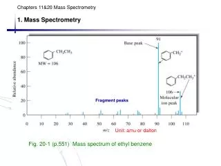
MOLECULAR MASS SPECTRA Ethyl benzene, molecular mass of 106 dalton. The analyte vapor was bombarded with a stream of electrons that led to the loss of an electron by the analyte and formation of the molecular ion M+ C6H5CH2CH3 + e- C6H5CH2CH.+3 + 2e- The charged species C6H5CH2CH.+3 is the molecular ion. Relaxation occurs by fragmentation of part of the molecular ions to produce ions of lower masses. The positive ions produced on electron impact are attracted through the slit of a mass spectrometer where they are sorted according to their mass-to-charge ratios and displayed in the form of a mass spectrum. The largest peak, termed the base peak.
ION SOURCES The starting point for a mass spectrometric analysis is the formation of gaseous analyte ions. Ion sources fall into two major categories: gas-phase sources and desorption sources. In the former, the sample is first vaporized and then ionized. In the latter, the sample in a solid or liquid state is converted directly into gaseous ions.
ION SOURCES Ion sources are also classified as being hard sources and soft sources. Hard sources impart sufficient energy to analyte molecules, relaxation involves rupture of bonds, producing fragment ions that have mass-to-charge ratios less than that of the molecular ion. Soft sources cause little fragmentation. Consequently, the resulting mass spectrum often consists of the molecular ion peak and only a few, if any other, peaks.
The Electron-Impact Sources The sample is brought to a temperature high enough to produce a molecular vapor, which is then ionized by bombarding the resulting molecules with a beam of energetic electrons. M + e- M.+ + 2e- where, M = analyte molecule, M.+ = molecular ion. Relaxation then usually takes place by extensive fragmentation, giving a large number of positive ions of various masses that are less than that of the molecular ion. These lower mass ions are called daughter ions.
Isotope Peaks Sometimes peaks occur at masses that are greater than that of the molecular ion. These peaks are attributable to ions having the same chemical formula but different isotopic compositions. e.g., for methylene chloride, the more important isotopic species are 12C1H235Cl2 (m = 84), 13C1H235Cl2 (m = 85), 12C1H235Cl37Cl (m = 86), 13C1H235Cl37Cl (m = 87), 12C1H237Cl2 (m = 88). The size of the various peaks depends upon the relative natural abundance of the isotopes.
Advantages and Disadvantages of El Sources Electron–impact sources are convenient to use because of good sensitivities. The extensive fragmentation and consequent large number of peaks is also an advantage because it often makes unambiguous identification of analytes possible. This fragmentation can also be a disadvantage, however, when it results in the disappearance of the molecular ion peak so that the molecular weight of analytes cannot be established. Another limitation of the electron-impact source is the need to volatilize the sample, which may result in thermal degradation of some analytes before ionization can occur. Electron-impact sources are only applicable to analytes having molecular weights smaller than about 103 daltons.
Chemical Ionization Sources In chemical ionization, gaseous atoms of the sample are ionized by collision with ions produced by electron bombardment of an excess of a reagent gas. A gaseous reagent is introduced into the ionization region in an amount such that the concentration ratio of reagent to sample is 103 to 104. Because of this large concentration difference the electron beam reacts nearly exclusively with reagent molecules.
Chemical Ionization Sources One of the most common reagents is methane which reacts with high-energy electrons to give several ions such as CH+4, CH+3, and CH+2. These ions react rapidly with additional methane molecules as follows: CH4+ + CH4 CH5++ CH3 CH3+ + CH4 C2H5++ H2
Chemical Ionization Sources Collisions between the sample molecule MH and CH5+ or C2H5+ are highly reactive and involve proton or hydride transfer. e.g., CH5+ + MH MH2++ CH4Proton transfer C2H5+ + MH MH2++ C2H4Proton transfer C2H5+ + MH M++ C2H6Hydride transfer Proton transfer reaction give the (M + 1)+ ion whereas the hydride transfer produces an ion with a mass one less than the analyte, or the (M - 1)+ ion. With some compounds, an (M + 29)+ peak is also produced from transfer of a C2H5+ ion to the analyte.
Instrument Components The block diagram in Fig. 20-11 shows the major components of mass spectrometers. The purpose of the inlet system is to introduce a very small amount of sample into the mass spectrometer, where its components are converted to gaseous ions. Ion sources of mass spectrometers convert the components of a sample into ions. The function of the mass analyzer is analogous to that of the grating in an optical spectrometer.
Instrument Components In the former, however, dispersion is based upon the mass-to-charge ratios of the analyte ions rather than upon the wavelength of photons. A mass spectrometer contains a transducer (for ions) that converts the beam of ions into an electrical signal that can then be processed, stored in the memory of a computer, and displayed or recorded in a variety of ways.