Download
1 / 24
240 likes | 245 Views
Chapter Goals. After studying this chapter, students should be able to . . . 1. describe the general functions of the major components of the heart. 2. describe the path of the blood through the heart and the function of the atrioventricular and semilunar valves.

E N D
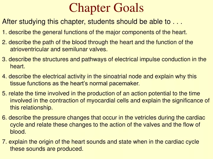
Chapter Goals After studying this chapter, students should be able to . . . 1. describe the general functions of the major components of the heart. 2. describe the path of the blood through the heart and the function of the atrioventricular and semilunar valves. 3. describe the structures and pathways of electrical impulse conduction in the heart. 4. describe the electrical activity in the sinoatrial node and explain why this tissue functions as the heart’s normal pacemaker. 5. relate the time involved in the production of an action potential to the time involved in the contraction of myocardial cells and explain the significance of this relationship. 6. describe the pressure changes that occur in the vetricles during the cardiac cycle and relate these changes to the action of the valves and the flow of blood. 7. explain the origin of the heart sounds and state when in the cardiac cycle these sounds are produced.
Chapter Goals 8. explain the cause of each wave in an electrocardiogram and relate these waves to other events in the cardiac cycle. 9. compare the structure of an artery and vein, and explain how the structure of each type of vessel relates to its function. 10. describe the structure of capillaries and explain the physiological significance of this structure. 11. define ischemia and discuss the possible causes of myocardial ischemia. 12. describe some common arrhythmias that can be detected with an ECG.
The Heart • Development • Structure • Valves • Electrical Activity • Cardiac Cycle
Anatomy 9-4
Cardiac Cycle • Phases • Systole • Diastole • Changes • Pressure • Electrical • Mechanical • Sound
Cardiac Control • a. Sympathetic - Increases heart rate, force of beating, and cardiac output (see below). (via norepinephrine) • b. Parasympathetic - opposite effect (via acetylcholine)
Chapter Summary Structure of the Heart I. The right and left sides of the heart pump blood through the pulmonary and systemic circulations. A. The right ventricle pumps blood to the lungs. This blood then returns to the left atrium. B. The left ventricle pumps blood into the aorta and systemic arteries. This blood then returns to the right atrium. II. The heart contains two pairs of one-way valves. A. The atrioventricular valves allow blood to flow from the atria to the ventricles, but not in the reverse direction. B. The semilunar valves allow blood to leave the ventricles and enter the pulmonary and systemic circulations, but these valves prevent blood from returning from the arteries to the ventricles.
Chapter Summary III. The electrical impulse begins in the sinoatrial node and spreads through both atria by electrical conduction from one myocardial cell to another. A. The impulse then excites the atrioventricular node, from which it is conducted by the bundle of His into the ventricles. B. The Purkinje fibers transmit the impulse into the ventricular muscle and cause it to contract.
Chapter Summary Cardiac Cycle and Heart Sounds I. The heart is a two-step pump. The atria contract first, and then the ventricles. A. During diastole, first the atria and then the ventricles fill with blood. B. The ventricles are about 80% filled before the atria contract and add the final 20% to the end-diastolic volume. C. Contraction of the ventricles ejects about two-thirds of their blood, leaving about one-third as the end-systolic volume. II. When the ventricles contract at systole, the pressure within them first rises sufficiently to close the AV valves and then rises sufficiently to open the semilunar valves. A. Blood is ejected from the ventricles until the pressure within the falls below the pressure in the arteries. At this point, the semilunar valves close and the ventricles begin relaxation. B. When the pressure in the ventricles falls below the pressure in the atria, a phase of rapid filling of the ventricles occurs, followed by the final filling caused by contraction of the atria.
Chapter Summary III. Closing of the AV valves produces the first heart sound, or "lub", at systole. Closing of the semilunar valves produces the second heart sound, or "dub". at diastole. Abnormal valves can cause abnormal sounds called murmurs.
Chapter Summary Electrical Activity of the Heart and the Electrocardiogram I. In the normal heart the impulse originates in the SA node, due to a spontaneous depolarization called the pacemaker potential. A. When this spontaneous depolarization reaches a threshold value, opening of the voltage-regulated Na+ gates and fast Ca2+ channels produces an action potential. B. Repolarization is produced by the outward diffusion of K+, but a stable resting membrane potential is not attained because spontaneous depolarization once more occurs. C. Other myocardial cells are capable of spontaneous activity, but the SA node is the normal pacemaker because its rate of spontaneous depolarization is the fastest. D. When the action potential produced by the SA node reaches other myocardial cells, they produce action potentials with a long plateau phase because of the slow, inward diffusion of Ca2+. E. The long action potential and long refractory period of myocardial cells allows the entire mass of cells to be in a refractory period while it contracts. This prevents the myocardium from being stimulated again until after it relaxes.
Chapter Summary II. The regular pattern of conduction in the heart produces a changing pattern of potential differences between two points on the body surface. A. The recording of this changing pattern caused by the heart’s electrical activity of the heart is called an electrocardiogram (ECG). B. The P wave is caused by depolarization of the atria; the QRS wave is caused by depolarization of the ventricles; and the T wave is produced by repolarization of the ventricles.