Download
1 / 18
190 likes | 415 Views
Organ systems in mammals. Table 40.1. Gas Exchange Among Different Animals. Respiration is the exchange of respiratory gases, oxygen and carbon dioxide, between the external environment and the cell or body. Occurs passively by diffusion

E N D
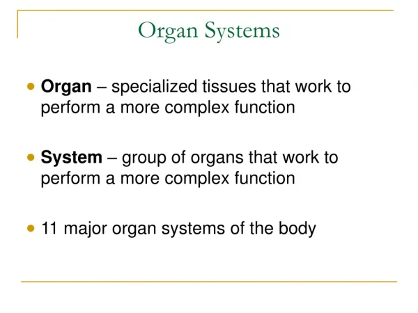
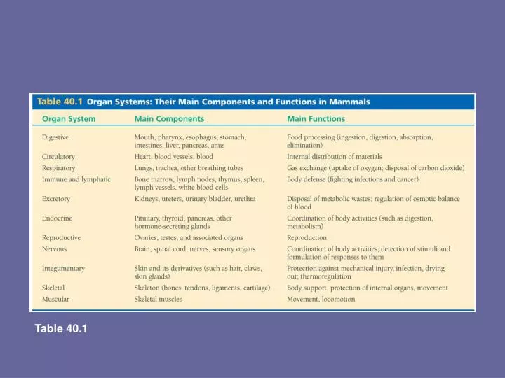
Organ systems in mammals Table 40.1
Gas Exchange Among Different Animals • Respiration is the exchange of respiratory gases, oxygen and carbon dioxide, between the external environment and the cell or body. • Occurs passively by diffusion • Respiratory surfaces must be thin, moist, and have a large surface area.
Respiratorymedium(air of water) O2 CO2 Respiratorysurface Organismal level Circulatory system Cellular level Energy-richmoleculesfrom food ATP Cellular respiration Figure 42.19 Concept 42.5: Gas exchange occurs across specialized respiratory surfaces • Gas exchange • Supplies oxygen for cellular respiration and disposes of carbon dioxide
Methods of Gas Exchange • Sponges and Hydra – gas exchange occurs over the entire surface of the organism wherever cells are in direct contact with the environment. • Earthworms and Flatworms – external respiratory surface, diffusion of oxygen and carbon dioxide occurs at the skin. Oxygen is carried by hemoglobin in the blood. • Grasshopper, other arthropods and crustaceans – internal respiratory surface, air enters through spiracles and travels through a system of tracheal tubes inot the body where diffusion occurs. Oxygen is carried by hemocyanin (uses copper instead of iron) • Fish – gills take advantage of countercurrent exchange to maximize the diffusion of respiratory gases
Air sacs Tracheae Spiracle (a) The respiratory system of an insect consists of branched internal tubes that deliver air directly to body cells. Rings of chitin reinforce the largest tubes, called tracheae, keeping them from collapsing. Enlarged portions of tracheae form air sacs near organs that require a large supply of oxygen. Air enters the tracheae through openings called spiracles on the insect’s body surface and passes into smaller tubes called tracheoles. The tracheoles are closed and contain fluid (blue-gray). When the animal is active and is using more O2, most of the fluid is withdrawn into the body. This increases the surface area of air in contact with cells. Figure 42.22a Tracheal Systems in Insects • Consists of tiny branching tubes that penetrate the body
Body cell Airsac Tracheole Body wall Myofibrils Trachea Air Tracheoles Mitochondria (b) This micrograph shows cross sections of tracheoles in a tiny piece of insect flight muscle (TEM). Each of the numerous mitochondria in the muscle cells lies within about 5 µm of a tracheole. Figure 42.22b 2.5 µm • The tracheal tubes • Supply O2 directly to body cells
Oxygen-poorblood Gill arch Oxygen-richblood Lamella Blood vessel Gill arch 15% 40% 70% 5% Water flow 30% Operculum 100% 60% 90% O2 Blood flowthrough capillariesin lamellaeshowing % O2 Water flowover lamellaeshowing % O2 Gillfilaments Figure 42.21 Countercurrent exchange The effectiveness of gas exchange in some gills, including those of fishes • Is increased by ventilation and countercurrent flow of blood and water
Gas Exchange in Humans • Air enters the nasal cavity and is moistened, warmed, and filtered. • Air passes through the larynx and down the trachea and bronchi and into the tiniest bronchioles, which end in microscopic air sacs called alveoli. • Diffusion of gases occurs at the alveoli
Branch from thepulmonaryartery(oxygen-poor blood) Branch from the pulmonary vein (oxygen-rich blood) Terminal bronchiole Nasalcavity Alveoli Pharynx Left lung Esophagus Larynx 50 µm Trachea 50 µm Right lung Bronchus Bronchiole Colorized SEM SEM Diaphragm Heart Figure 42.23 The Human Respiratory System Branching ducts deliver oxygen to the lungs
Concept 42.6: Breathing ventilates the lungs • Breathing is the alternate inhalation and exhalation of air. • An amphibian such as a frog • Ventilates its lungs by positive pressure breathing, which forces air down the trachea.
Rib cage expands asrib muscles contract Rib cage gets smaller asrib muscles relax Air inhaled Air exhaled Lung Diaphragm INHALATIONDiaphragm contracts(moves down) EXHALATIONDiaphragm relaxes(moves up) Figure 42.24 Mammals ventilate their lungs • By negative pressure breathing, which pulls air into the lungs
Cerebrospinalfluid The medulla’s control center also helps regulate blood CO2 level. Sensorsin the medulla detect changes in the pH (reflecting CO2 concentration) of the blood and cerebrospinal fluid bathing the surface of the brain. 1 The control center in the medulla sets the basicrhythm, and a control centerin the pons moderates it,smoothing out thetransitions between inhalations and exhalations. 5 Nerve impulses relay changes in CO2 and O2 concentrations. Other sensors in the walls of the aortaand carotid arteries in the neck detect changes in blood pH andsend nerve impulses to the medulla. In response, the medulla’s breathingcontrol center alters the rate anddepth of breathing, increasing bothto dispose of excess CO2 or decreasingboth if CO2 levels are depressed. Pons Breathing control centers Nerve impulses trigger muscle contraction. Nervesfrom a breathing control centerin the medulla oblongata of the brain send impulses to thediaphragm and rib muscles, stimulating them to contractand causing inhalation. 2 Medullaoblongata Carotidarteries Aorta In a person at rest, these nerve impulses result in about 10 to 14 inhalationsper minute. Between inhalations, the musclesrelax and the person exhales. 3 The sensors in the aorta andcarotid arteries also detect changesin O2 levels in the blood and signal the medulla to increase the breathing rate when levels become very low. 6 Figure 42.26 Diaphragm Rib muscles Control of Breathing in Humans • The main breathing control centers • Are located in two regions of the brain, the medulla oblongata and the pons 4
The centers in the medulla • Regulate the rate and depth of breathing in response to pH changes in the cerebrospinal fluid • The medulla adjusts breathing rate and depth • To match metabolic demands • Sensors in the aorta and carotid arteries • Monitor O2 and CO2 concentrations in the blood • Exert secondary control over breathing
Heme group Iron atom O2 loaded in lungs O2 O2 unloaded In tissues O2 Polypeptide chain Hemoglobin • Like all respiratory pigments • Hemoglobin must reversibly bind O2, loading O2 in the lungs and unloading it in other parts of the body • Hemoglobin is an allosteric molecule and exhibits cooperativity. Binding of oxygen to one subunit increases affinity for the other subunits. • A drop in pH lowers the affinity of hemoglobin for oxygen (Bohr shift). Actively respiring tissue releases CO2 and lowers pH of the surrounding by forming carbonic acid in water. Figure 42.28
Factors Affecting the Dissociation Curve of Hemoglobin • At lower pH, the hemoglobin has less affinity for oxygen (the dissociation curve is further to the right). • Organisms with higher metabolism have a greater need for oxygen and their hemoglobin has a lower affinity for oxygen (the dissociation curve of the organism with the higher metabolism is to the right) • Fetal hemoglobin has a higher affinity for oxygen than adult hemoglobin so it can take oxygen from the maternal hemoglobin. • Mammals that evolved at high altitudes must have hemoglobin with a greater affinity for oxygen because less oxygen is available there (curve is to the left).
(a) PO2 and Hemoglobin Dissociation at 37°C and pH 7.4 O2 unloaded from hemoglobin during normal metabolism 100 80 O2 reserve that can be unloaded from hemoglobin to tissues with high metabolism 60 O2 saturation of hemoglobin (%) 40 20 0 60 100 40 80 0 20 Tissues at rest Lungs Tissues during exercise PO2 (mm Hg) (b) pH and Hemoglobin Dissociation 100 pH 7.4 80 Bohr shift:Additional O2released from hemoglobin at lower pH(higher CO2concentration) 60 O2 saturation of hemoglobin (%) pH 7.2 40 20 0 Figure 42.29a, b 60 100 40 80 0 20 PO2 (mm Hg)
Transport of Carbon Dioxide • A small amount of carbon dioxide is transported by hemoglobin • Most carbon dioxide is carried in the blood as part of the carbonic acid-bicarbonate ion system, which maintains the blood at a constant pH 7.4. • Carbon dioxide combines with water to form carbonic acid (catalyzed by carbonic anhydrase). • Carbonic acid dissociates into bicarbonate ion and a proton. The protons can be given up into the plasma, which lowers the blood pH, or taken up by bicarbonate ion, which raises the blood pH. Equation: CO2 + H2O H2CO3 + H2CO3- + H+
Tissue cell Carbon dioxide produced bybody tissues diffuses into the interstitial fluid and the plasma. Most of the HCO3– diffuseinto the plasma where it is carried in the bloodstream to the lungs. CO2 transportfrom tissues 11 10 7 6 5 4 3 2 1 8 9 CO2 produced Interstitialfluid CO2 Over 90% of the CO2 diffuses into red blood cells, leaving only 7%in the plasma as dissolved CO2. Blood plasmawithin capillary CO2 Capillarywall In the HCO3– diffusefrom the plasma red blood cells, combining with H+ released from hemoglobin and forming H2CO3. CO2 H2O Some CO2 is picked up and transported by hemoglobin. Redbloodcell Hemoglobinpicks upCO2 and H+ H2CO3 Hb Carbonic acid Carbonic acid is converted back into CO2 and water. HCO3– + H+ Bicarbonate However, most CO2 reacts with water in red blood cells, forming carbonic acid (H2CO3), a reaction catalyzed bycarbonic anhydrase contained. Withinred blood cells. HCO3– To lungs CO2 formed from H2CO3 is unloadedfrom hemoglobin and diffuses into the interstitial fluid. CO2 transportto lungs HCO3– 9 6 2 7 5 4 3 1 8 + H+ HCO3– CO2 diffuses into the alveolarspace, from which it is expelledduring exhalation. The reductionof CO2 concentration in the plasmadrives the breakdown of H2CO3 Into CO2 and water in the red bloodcells (see step 9), a reversal of the reaction that occurs in the tissues (see step 4). Carbonic acid dissociates into a biocarbonate ion (HCO3–) and a hydrogen ion (H+). HemoglobinreleasesCO2 and H+ Hb H2CO3 H2O CO2 Hemoglobin binds most of the H+ from H2CO3 preventing the H+from acidifying the blood and thuspreventing the Bohr shift. CO2 11 10 CO2 CO2 Figure 42.30 Alveolar space in lung