Download
1 / 1
10 likes | 149 Views
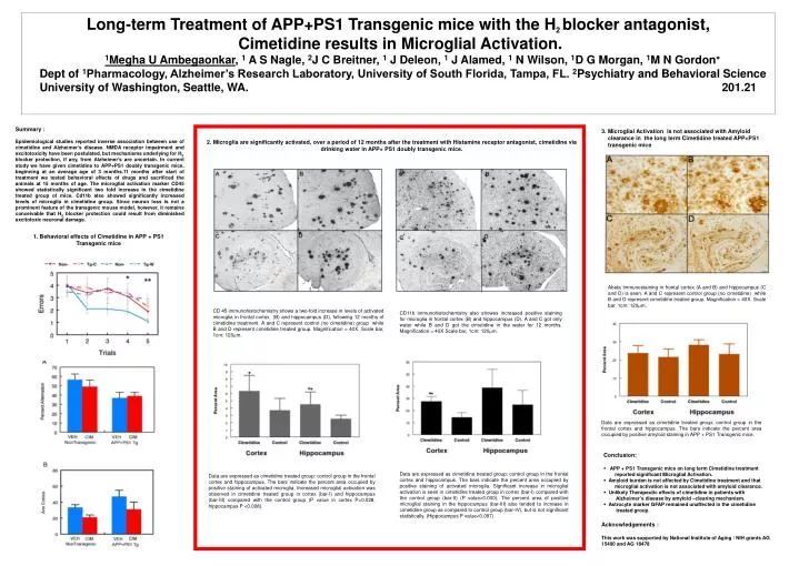
Long-term Treatment of APP+PS1 Transgenic mice with the H 2 blocker antagonist, Cimetidine results in Microglial Activation. 1 Megha U Ambegaonkar , 1 A S Nagle, 2 J C Breitner, 1 J Deleon, 1 J Alamed, 1 N Wilson, 1 D G Morgan, 1 M N Gordon

E N D
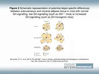
Long-term Treatment of APP+PS1 Transgenic mice with the H2 blocker antagonist, Cimetidine results in Microglial Activation. 1Megha U Ambegaonkar, 1 A S Nagle, 2J C Breitner, 1 J Deleon, 1 J Alamed, 1 N Wilson, 1D G Morgan, 1M N Gordon Dept of 1Pharmacology, Alzheimer’s Research Laboratory, University of South Florida, Tampa, FL. 2Psychiatry and Behavioral Science University of Washington, Seattle, WA. 201.21 Summary : Epidemiological studies reported inverse association between use of cimetidine and Alzheimer’s disease. NMDA receptor impairment and excitotoxicity have been postulated, but mechanisms underlying for H2 blocker protection, if any, from Alzheimer’s are uncertain. In current study we have given cimetidine to APP+PS1 doubly transgenic mice, beginning at an average age of 3 months.11 months after start of treatment we tested behavioral effects of drugs and sacrificed the animals at 15 months of age. The microglial activation marker CD45 showed statistically significant two fold increase in the cimetidine treated group of mice. Cd11b also showed significantly increased levels of microglia in cimetidine group. Since neuron loss is not a prominent feature of the transgenic mouse model, however, it remains conceivable that H2 blocker protection could result from diminished excitotoxic neuronal damage. 3. Microglial Activation is not associated with Amyloid clearance in the long term Cimetidine treated APP+PS1 transgenic mice 2. Microglia are significantly activated, over a period of 12 months after the treatment with Histamine receptor antagonist, cimetidine via drinking water in APP+ PS1 doubly transgenic mice. 1. Behavioral effects of Cimetidine in APP + PS1 Transgenic mice Abeta Immunostaining in frontal cortex (A and B) and hippocampus (C and D) is seen. A and C represent control group (no cimetidine) while B and D represent cimetidine treated group. Magnification = 40X. Scale bar, 1cm: 120m. CD 45 immunohistochemistry shows a two-fold increase in levels of activated microglia in frontal cortex (B) and hippocampus (D), following 12 months of cimetidine treatment. A and C represent control (no cimetidine) group while B and D represent cimetidine treated group. Magnification = 40X. Scale bar, 1cm: 120m. CD11b immunohistochemistry also showes increased positive staining for microglia in frontal cortex (B) and hippocampus (D). A and C got only water while B and D got the cimetidine in the water for 12 months. Magnification = 40X.Scale bar, 1cm: 120m. Data are expressed as cimetidine treated group: control group in the frontal cortex and hippocampus. The bars indicate the percent area occupied by positive amyloid staining in APP + PS1 Transgenic mice. Conclusion: • APP + PS1 Transgenic mice on long term Cimetidine treatment reported significant Microglial Activation. • Amyloid burden is not affected by Cimetidine treatment and that microglial activation is not associated with amyloid clearance. • Unlikely Therapeutic effects of cimetidine in patients with Alzheimer’s disease by amyloid –clearing mechanism. • Astrocyte marker GFAP remained unaffected in the cimetidine treated group. Data are expressed as cimetidine treated group: control group in the frontal cortex and hippocampus. The bars indicate the percent area occupied by positive staining of activated microglia. Significant increase in microglial activation is seen in cimetidine treated group in cortex (bar-I) compared with the control group (bar-II) (P value<0.000). The percent area of positive microglial staining in the hippocampus (bar-III) also tended to increase in cimetidine group as compared to control group (bar-IV), but is not significant statistically. (Hippocampus P value<0.087) Data are expressed as cimetidine treated group: control group in the frontal cortex and hippocampus. The bars indicate the percent area occupied by positive staining of activated microglia. Increased microglial activation was observed in cimetidine treated group in cortex (bar-I) and hippocampus (bar-III) compared with the control group (P value in cortex P<0.028, hippocampus P <0.008). Acknowledgements : This work was supported by National Institute of Aging / NIH grants AG 15490 and AG 18478