Download
1 / 20
200 likes | 367 Views
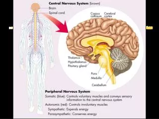
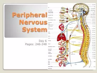
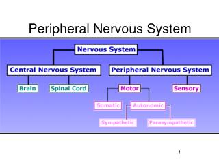
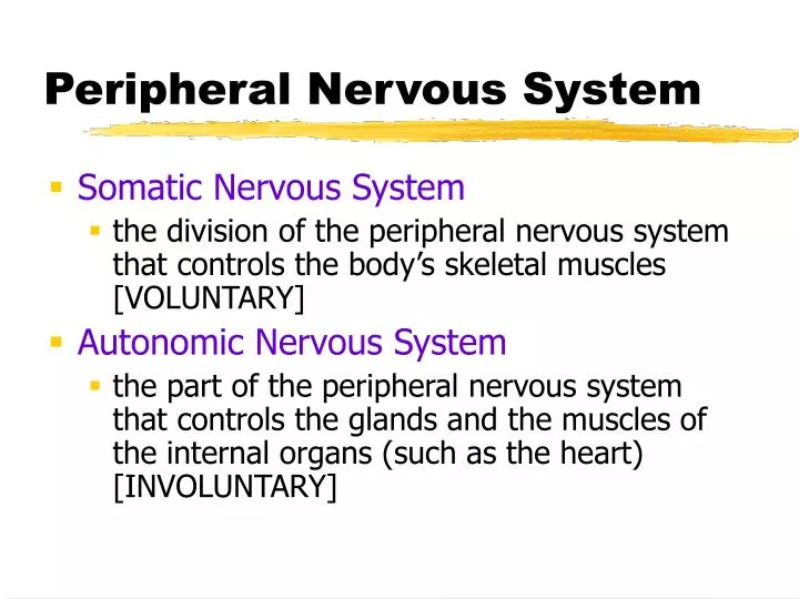
Peripheral Nervous System. Somatic Nervous System the division of the peripheral nervous system that controls the body’s skeletal muscles [VOLUNTARY] Autonomic Nervous System

E N D
Peripheral Nervous System • Somatic Nervous System • the division of the peripheral nervous system that controls the body’s skeletal muscles [VOLUNTARY] • Autonomic Nervous System • the part of the peripheral nervous system that controls the glands and the muscles of the internal organs (such as the heart) [INVOLUNTARY]
Autonomic Nervous System • Sympathetic Nervous System • division of the autonomic nervous system that AROUSES the body, mobilizing its energy in stressful situations • Parasympathetic Nervous System • division of the autonomic nervous system that CALMS the body, conserving its energy
The Brain • Brainstem • the oldest part and central core of the brain, beginning where the spinal cord swells as it enters the skull • responsible for automatic survival functions
The Brainstem • Medulla [muh-DUL-uh] • base of the brainstem • controls heartbeat and breathing • Pons • Connects different brain regions together • Involved in facial expressions
The Brainstem • Reticular Formation • a nerve network in the brainstem that plays an important role in controlling arousal • Thalamus [THAL-uh-muss] • the brain’s sensory switchboard, located on top of the brainstem • it directs messages to the sensory receiving areas in the cortex and transmits replies to the cerebellumand medulla
The Brainstem • Cerebellum [sehr-uh-BELL-um] • the “little brain” attached to the rear of the brainstem • it helps coordinate voluntary movement and balance • Formulates implicit memories
The Limbic System • a doughnut-shaped system of neural structures at the border of the brainstem and cerebral hemispheres • associated with emotions such as fear and aggression and drives such as those for food and sex • includes the hippocampus, amygdala, and hypothalamus.
The Limbic System • Amygdala [ah-MIG-dah-la] • two almond-shaped neural clusters that are components of the limbic system and are linked to emotion (specifically aggression and fear)
The Limbic System • Hypothalamus • neural structure lying below (hypo) the thalamus; directs several maintenance activities • Eating and drinking • Sex drive • body temperature • helps govern the endocrine system via the pituitary gland
Hypothalamus Stimulation • Electrode implanted in reward center of hypothalamus • Rat readily crosses to get stimulation
The Limbic System • Hippocampus: a structure in the limbic system linked to explicit memory (Clive Wearing example) • Pituitary Gland: master endocrine gland, linked to growth (Andre theGiant example)
The Cerebral Cortex Laid out it would be about the size of a large pizza. • Cerebral Cortex • the intricate fabric of interconnected neural cells that covers the cerebral hemispheres
The Cerebral Cortex • Frontal Lobes • involved in speaking and muscle movements and in making plans and judgments (Phineas Gage Clip) • Parietal Lobes • Involved in sensations (touch), pressure, and pain
The Cerebral Cortex • Occipital Lobes • include the visual areas, which receive visual information from the opposite visual field • Temporal Lobes • include the auditory areas
The Cerebral Cortex • Motor Cortex • area at the rear of the frontal lobes that controls voluntary movements • Sensory Cortex • area at the front of the parietal lobes that registers and processes body sensations
The Cerebral Cortex • Functional MRI scan shows the visual cortex activated as the subject looks at faces
Association Areas • “Uncommitted” areas that are not involved in primary functions but play a role in learning, remembering, and thinking • More intelligent animals have increased “uncommitted” or association areas of the cortex