Download
1 / 20
200 likes | 345 Views
Non-Atherosclerotic Arterial disease-Cerebral. Hemorrhage: Underlying cause of 16% of all strokes. Hypertension is leading cause of cerebral hemorrhage. Aneurysm: can be congenital or result from atherosclerosis. Hemorrhage cont. Trauma can cause intracerebral or subarachnoid bleed.

E N D
Non-Atherosclerotic Arterial disease-Cerebral • Hemorrhage: Underlying cause of 16% of all strokes. • Hypertension is leading cause of cerebral hemorrhage. • Aneurysm: can be congenital or result from atherosclerosis.
Hemorrhage cont. • Trauma can cause intracerebral or subarachnoid bleed. • Thrombolytic Therapy: Lysis of thrombus (dissolution of or decomposition of thrombus) • Heparin Therapy: anticoagulant
Non-athero cerebral: • Emboli • Greatest number of cerebral emboli come from the heart. • Most go to the posterior circ • Numerous materials can embolize. • Air • Tumor • Thrombus • atherosclerosis
Cerebral cont. • Aneurysm • Congenital: Berry (small, sacular), Arterio-venous malformation (avm) artery and vein connected causing shunting of flow. • Hypertension • Trauma (AVM)
Non-athero cerebral cont. • Inflammatory conditions: • Sickle Cell: young, African-American population • Periarteritis Nodosa/Polyarteritis: • A collegen / allergic disease • Necrosis of media and thickening of the intima • Segmental arteritis and possible small aneurysms occur • Can obstruct function of any arterial system involved
Cerebral cont. • Temporal Arteritis • Intimal proliferation and inflammation/cause unknown/ because patients also have polymyalgia rheumatica it is considered a rheumatic disease. • It affects medium size branches of carotid arteries, coronaries, aorta and it’s branches. • Complications: blindness, stroke, heart attack, and norrowing of major aortic branches. • 50 YO women. Women>Men.
Non-athero extracranial causes . • Takayasu’s: also a form of arteritis and is distinguished from Temporal Arteritis microscopically and clinically. • Predominantly affects young women,Asian. • Involves predominantly the aortic arch and it’s branches. Death is caused by CHF and CVA • Treated with corticosteroids
Non-athero extracranial causes cont. • Periarteritis nodosa/ polyarteritis • An inflammatory disease of small and medium sized arteries. Affects any organ or body system. • Temporal Arteritis • Reference: Cardiology Clinics PVD in the Elderly. 8-1991. • Reference: Diseases of the Heart and Circulation. 3rd ed. Wood, Paul page.727
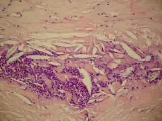
Non-athero extracranial cont. • Carotid Body Tumor • Carotid body: 1X1 mm, in adventitia at carotid bifurcation, a component of the autonomic nervous system that helps control arterial PH, blood gas level, and blood pressure. • A paraganglioma, low incidence of malignance, highly vascular. • Located most often between ECA and ICA. • Causes palpable mass, neck pain, headache, laryngeal nerve palsy, and invasion of carotid arteries.
Non-athero causes extracranial • Dissection: Under forced pressure blood separates layers of the arterial wall. • Usually trauma • Can be spontaneous • Spontaneous dissection many times results with nonviolent trauma: exercise or rapid neck motion. • Contributing factors to spontaneous dissection: hypertension, fibromuscular hyperplasia, and conditions that weaken the arterial wall – Marfan’s syndrome, cystic medial necrosis, and Ehlers-Danlos syndrome.
Dissection cont. • Thrombosing is associated • Occlusion or hemdynamic stenosis • Embolic source • Either can cause TIA/CVA • Anitcoagulate or thrombectomy and repair wall
Dissection cont. • A false lumen is created. • If separation is between media and adventia a pseudoaneurysm can occur.
Non-athero renal pathologies • Fibromuscular Dysplasia: Hyperplasia of the media or intimal layer of the renal artery. Forms concentric bands usually located in the mid to distal renal arteries. • Occurs more frequently in women • Onset can be at early age (teens) or before 50. • More than one band forms creating “tandem lesions). Known as “string of beads” on arteriogram.
Non-athero renal pathologies • Rare causes of renovascular disease • Takayasu’s/ Polyarteritis • Renal artery thrombosis or embolism • Extrinsic renal artery compression by cyst or tumor • Abdominal aortic coarctation • Congenital vascular lesions • Reference: Bernstein, 4th ed, page 652
Non-atherosclerotic mesenteric lesions • Compression syndrome • Median Arcuate ligament can compression the lumen of the celiac trunk or SMA • This is most often intermittant • Rarely a cause of bowel ischemia
Non-athero mesenteric lesions cont. • Emboli • Most often from heart • Thrombus • Tumor
References Vas Phy 2 • Slide 1 Handout from Bowman Grey lecture on carotid duplex, 1984. • Slide 2 Cardiology Clinics, PVD in Elderly, August 1991, Saunders. Breslin, Ed. Pgs.508-509 • Slides 5 & 7 Taber’s Cyclopedic Medical Dictionary, Davis, 1985. • Slide 6 Diseases of the Heart and Circulation 3rd ed. Wood’s, Paul. Lippincott, 1969.pg 727.
Refer Vas Phy 2 cont. • Slides 7,8,9 Cardiology Clinics, PVD in Elderly, August 1991. PGS 547-553 • Slide 10 Introductin to Vascular Ultrasound, 4th Ed., Zwiebel, Saunders, 2000.pgs. 163-165 &160. • Slides 11,12,13 Cardiology Clinics, PVD in Elderly, August 1991. Pg.528. / Introduction to Vascular Ultrasound,4th Ed. Pages 156-161.
Ref Vas Physi 2 cont. • Slide 14 Cardiology Clinics, PVD in Elderly, August 1991. Pgs 528-530./ Ciba, Heart vol 5. 1981, Pg 229. • Slide 15 Vascular Diagnosis, 4th Ed., Bernstein, Mosby, 1993, Pg 652 • Slide 16 Introduction to Vascular Ultrasonography, Zwiebel 4th, Ed. Saunders,2000 Pgs 421-422.