Download
1 / 1

E N D
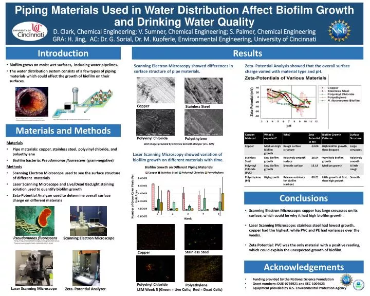
Piping Materials Used in Water Distribution Affect Biofilm Growth and Drinking Water QualityD. Clark, Chemical Engineering; V. Sumner, Chemical Engineering; S. Palmer, Chemical Engineering GRA: H. Jing, AC: Dr. G. Sorial, Dr. M. Kupferle, Environmental Engineering, University of Cincinnati Results Introduction • Biofilm grows on moist wet surfaces, including water pipelines. • The water distribution system consists of a few types of piping materials which could effect the growth of biofilm on their surfaces. Scanning Electron Microscopy showed differences in surface structure of pipe materials. Zeta–Potential Analysis showed that the overall surface charge varied with material type and pH. Stainless Steel Copper http://www.premierwatermn.com/water-quality/water-contaminants/bacteria-virus-and-microorganisms-in-water/ http://water.epa.gov/lawsregs/rulesregs/sdwa/tcr/distributionsystems.cfm Copper Stainless Steel Materials and Methods • Materials • Pipe materials: copper, stainless steel, polyvinyl chloride, and polyethylene • Biofilm bacteria: Pseudomonas fluorescens (gram-negative) • Methods • Scanning Electron Microscope used to see the surface structure of different materials • Laser Scanning Microscope and Live/Dead BacLight staining solution used to quantify biofilm growth • Zeta-Potential Analyzer used to determine overall surface charge on different materials SEM images provided by Christina Bennett-Stamper (U.S. EPA) Polyvinyl Chloride Polyethylene Laser Scanning Microscopy showed variation of biofilm growth on different materials with time. Polyvinyl Chloride Polyethylene Conclusions • Scanning Electron Microscope: copper has large crevasses on its surface, which could be why it had high biofilm growth. • Laser Scanning Microscope: stainless steel had lowest growth, copper had the highest, while PVC and PE had variances over the weeks. • Zeta Potential: PVC was the only material with a positive reading, which could explain the unexpected growth of biofilm. Pseudomonas fluorescens Scanning Electron Microscope • http://organicsoiltechnology.com/pseudomonas-fluorescens-phosphate-solubilization.html Acknowledgements • Funding provided by the National Science Foundation • Grant numbers: DUE-0756921 and EEC-1004623 • Equipment provided by U.S. Environmental Protection Agency LSM Week 5 (Green = Live Cells; Red = Dead Cells) Laser Scanning Microscope Zeta–Potential Analyzer