Download
1 / 24
300 likes | 602 Views
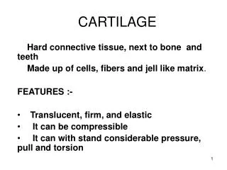
The biology of cartilage. Hyaline Cartilage. has a biomechanic function is localized on the articular surfaces of the joints. . Cartilage function.

E N D
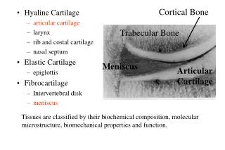
Hyaline Cartilage • has a biomechanic function • is localized on the articular surfaces of the joints.
Cartilage function • Shock absorbant: although it is at most only a few millimeters thick, it has surprising stiffness to compression and resilience; it also has an exceptional ability to distribute loads, thereby minimizing peak stresses on subchondral bone. • Most important characteristic: its durability. In most people it provides normal joint function for eighty years or more. No synthetic material approaches this level of performance.
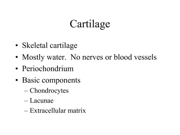
Cartilage histology • it’s a skeletal connective tissue • it has a mesodermal origin • it has a peculiar organization.
Cartilage histology Adult articular cartilage appears to be a simple inert tissue. Light microscopy shows that it consists primarily of ECM, with only one type of cell, the chondrocyte. It lacks blood vessels, lymphatic vessels and nerves. Compared with tissues such as muscle or bone, cartilage has a low level of metabolic activity and is less responsive to changes in loading and to injury.
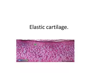
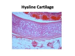
Cartilage histology • elasticcartilage • fibrous cartilage • hyaline cartilage
Cartilage histology • elastic cartilage • fibrous cartilage • hyaline cartilage
Cartilage histology • elastic cartilage • fibrous cartilage • hyaline cartilage
Hyaline cartilage components: • cellular component : chondrocytes (about 1% of the total volume) • extracellular matrix : collagen type II, HA, glycosaminoglycans, proteoglycans, water. As in other connective tissues, articular cartilage derives its form and mechanical properties from its matrix
The chondrocytes • Chondrocytes surround themselves with their ECM and do not form cell-to-cell contacts. • Chondrocyte characteristics: • Spheroidal shape • Synthesis of type II collagen, large aggregating proteoglycans and specific non-collagenous proteins • Formation of these molecules into the ECM. • High metabolic activity (the total metabolic activity of the tissue is low due to the low cell density)
The chondrocytes Maintenance of the articular surface requires turnover of the matrix macromolecules and alterations in the macromolecular framework of the matrix in response to use of the joint. To accomplish these activities, the cells must sense changes in the composition of the matrix that are due to degradation of macromolecules, as well as changes in the demands placed on the articular surface. This sensing function is probably performed by short cilia extending from the cell into the matrix.
The chondrocytes • In adult animals, chondrocytes derive their nutrition from nutrients in the synovial fluid, which, to reach the cell, must pass through a double diffusion barrier: • synovial tissue and synovial fluid • cartilage matrix. • The nature of this system leaves chondrocytes with a low concentration of oxygen relative to most other tissues; therefore, they depend primarily on anaerobic metabolism.
Collagen fibers Chondrocite Proteoglican The extracellular matrix
The extracellular matrix • It’s made of tissue fluid and the framework of structural macromolecules that give the tissue its form and stability. • Tissue fluid: • water: 80% of wet weight of articular cartilage • gases, small proteins, metabolites and a high concentration of cations to balance the negatively charged proteoglycans.
The extracellular matrix • Structural macromolecules (20-40% of the wet weight of the tissue): • collagens (60% of dry weight of cartilage) • proteoglycans (25-35% of dry weight) • glycoproteins (15-20% of dry weight) • Collagens are distributed uniformly throughout the depth of the cartilage and provide the tissue with its form and tensile strength. • Proteoglycans and glycoproteins bind to the collagen meshwork and help in keeping water trapped inside.
The collagens • Articular cartilage contains multiple genetically distinct collagen types: • type II • type VI • type IX • type X • type XI • Collagenstype II, IX and XI form the cross-banded fibrils seen with electron microscopy. • Collagen type II accounts for 90 to 95% of the collagen in articular cartilage.
The proteoglycans • Consist of a protein core and one or more glycosaminoglycan chains (long unbranched polysaccharide chains consisting of repeating disaccharides). • Two classes in articular cartilage: • Large aggregating proteoglycan monomers or aggrecans. • Small proteoglycans (decorin, fibromodulin, etc.)
Proteoglycan aggregates Most aggregans are linked with hyaluronic acid to form proteoglycan aggregates. These large molecules have a central backbone of hyaluronan that can range in length from several hundred to more than 10,000 nanometers. Large aggregates may have more than 300 associated aggrecan molecules.
Hyaluronan • It’s one of the critical molecules for the maintenance of the physico-chemicals characteristics of the extracellular matrix of articular cartilage.