Download
1 / 48
480 likes | 573 Views
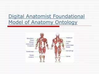
Digital Human Open Source Software Framework for Organ Modeling and Simulation. The Foundational Model of Anatomy Cornelius Rosse, M.D., D.Sc. Structural Informatics Group University of Washington. Organ Modeling and Simulation. Challenges Interoperability of software

E N D
Digital Human Open Source Software Framework for Organ Modeling and Simulation The Foundational Model of Anatomy Cornelius Rosse, M.D., D.Sc. Structural Informatics Group University of Washington
Organ Modeling and Simulation • Challenges • Interoperability of software • Representation of biomedical knowledge in machine-processible form • Integration and correlation of • biomedical information
The Semantic Web: A new form of Web content that is meaningful to computers will unleash a revolution of new possibilities. By: Tim Berners-Lee James Hendler Ora Lassila
Digital Anatomist Information Project • Motivation • Manifestations of health and disease are attributes of anatomical structures. • Logical and comprehensive representation of anatomical knowledge can serve as a foundation for other types of biomedical information.
Digital Anatomist Project University of Washington
End-User Programs Authoring Programs Network Servers Symbolic Knowledge Source Image Repository Anatomy Knowledge Sources Digital Anatomist Information System
End-User Programs Authoring Programs Servers Digital Anatomist Information System Network 2-D Images 2-D Annotations 3-D Model 3-D Image Volumes Symbolic Knowledge Source Image Repository
End-User Programs Servers Digital Anatomist Information System Symbolic Authoring Programs Foundational Model Builder Protégé Network Foundational Model Meta- knowledge Clinical Info Image Repository Symbolic Knowledge Sources
Organ Modeling and Simulation • What is an Organ? • Organ [L. organum; Gr. organon] • a somewhat independent part of the body that performs a special function or functions. • Dorland's Medical Dictionary • E.g., liver, heart, lung, kidney • hand, erythrocyte • Organ • in animals and plants, a part composed of several tissues and adopted to perform a specific function or functions. • Webster’s Dictionary • E.g., liver, heart, lung, kidney • hand, head of femur, right ventricle
Challenges of Anatomical Knowledge Representation Controlled Medical Terminologies (CMT) • MeSH (Medical Subject Headings) • SNOMED (Systematized Nomenclature of Medicine) • The Read Codes • GALEN (General Architecture for Languages • Encyclopedias and Nomenclatures in Medicine) • NeuroNames (University of Washington) • UMLS (Unified Medical Language Systems) • US National Library of Medicine
Challenges of Anatomical Knowledge Representation • Conclusion • Inadequacy of • traditional knowledge sources • New need for • computer-processable • anatomical knowledge
Challenges of Anatomical Knowledge Representation • Question: • What information should be entered in the • Foundational Model of Anatomy? • Answer: • The structure of anatomical structures • that constitute the body. • StructureBody= ({SubobjectBody}, {Structural relationship})
What kind of information? • “The oesophagus is a muscular tube … connecting the pharynx to the stomach. It begins in the neck, level with the lower border of the cricoid cartilage and the sixth cervical vertebra; descending largely anterior to the vertebral column through the superior and posterior mediastina.” • Gray’s Anatomy, 38th edition, p. 1751
What kind of information? • “The oesophagus is a muscular tube … connecting the pharynx to the stomach. It begins in the neck, level with the lower border of the cricoid cartilage and the sixth cervical vertebra; descending largely anterior to the vertebral column through the superior and posterior mediastina.” • Gray’s Anatomy, 38th edition, p. 1751
What kind of information? • “The oesophagusis amuscular tube … connecting the pharynx to the stomach. Itbeginsin the neck, level with the lower border of the cricoid cartilage and the sixth cervical vertebra; descending largely anteriorto the vertebral columnthrough the superior and posterior mediastina.” • Gray’s Anatomy, 38th edition, p. 1751
What kind of information? • Symbolic model, • a conceptualization of a domain of discourse • represented with non-graphical symbols; • in computer-processible (“understandable”) form; • supports inference (reasoning).
What is the Foundational Model of Anatomy (FM)? • Foundational Model of Anatomy • is a symbolic model of the physical organization of the human body; • declares the principles • for including concepts and relationships • that are implicitly assumed • when knowledge of anatomy • is applied in different contexts; • explicitly defines • concepts and relationships • necessary and sufficient for consistently • modeling the structure of the • human body.
Foundational Model of Anatomy Fm = (Ao, ASA, ATA, Mk) where: Ao = Anatomy ontology ASA = Anatomical Structural Abstraction ATA = Anatomical Transformation Abstraction Mk = Metaknowledge (principles, rules, axioms)
Foundational Model of Anatomy Anatomical Structural Abstraction Fm = (Ao, ASA, ATA, Mk) (1) ASA = (Do, Bn, Pn, SAn) (2) where: Do = Dimensional ontology Bn = Boundary network Pn = Part-of network SAn = Spatial Association network
Foundational Model of Anatomy Spatial Association Network Fm = (Ao, ASA, ATA, Mk) (1) ASA = (Do, Bn, Pn, SAn) (2) SAn = (Ln, On, Cn) (3) where: Ln = Location On = Orientation Cn = Connectivity
Networks of ASA Right Ventricle
Anatomy Ontology Anatomical Structure Organ Part Organ Subdivision Cardiac Chamber -is a- Right Ventricle
Dimensional Ontology Anatomy Ontology Anatomical Structure Volume (3-D) Organ Part Organ Subdivision Polyhedron Cardiac Chamber -is a- Right Ventricle
Spatial Ontology Anatomy Ontology Boundary Network Anatomical Structure Line (1-D) Surface (2-D) Volume (3-D) Organ Part Anterior Interventricular Sulcus Anatomical Surface Organ Subdivision Polyhedron -is a- Right Coronary Sulcus Cardiac Chamber Sternocostal Surface bounded by -is a- bounded by Inferior margin of heart boundary of Diaphragmatic Surface Coronary Sulcus bounded by Posterior IV Sulcus Anatomical Landmark -is a- Apex Crux of heart Point (1-D) Right Ventricle
Spatial Ontology Anatomy Ontology Boundary Network Anatomical Structure Line (1-D) Surface (2-D) Volume (3-D) Organ Part Anterior Interventricular Sulcus Anatomical Surface Organ Subdivision Polyhedron -is a- -is a- Heart Right Coronary Sulcus Cardiac Chamber Sternocostal Surface bounded by Part-of Network has -is a- super- object bounded by Infundibulum Inferior margin of heart Inflow part of RV has boundary of subobject Diaphragmatic Surface Coronary Sulcus Wall of RV Cavity of RV bounded by Posterior IV Sulcus Anatomical Landmark Cavity of infund. Cavity of infl.part -is a- Apex Crux of heart Point (1-D) Right Ventricle
Spatial Ontology Anatomy Ontology Boundary Network Anatomical Structure Line (1-D) Surface (2-D) Volume (3-D) Organ Part Anterior Interventricular Sulcus Anatomical Surface Organ Subdivision Polyhedron -is a- -is a- Heart Right Coronary Sulcus Cardiac Chamber Sternocostal Surface bounded by Part-of Network has -is a- super- object bounded by Infundibulum Inferior margin of heart Inflow part of RV has boundary of subobject Diaphragmatic Surface Coronary Sulcus Wall of RV has adjacency Cavity of RV bounded by has adjacency Posterior IV Sulcus Anatomical Landmark Cavity of infund. Cavity of infl.part to left anterior inferior inferior Left ventricle -is a- Pericardial sac Diaphragm Apex Crux of heart Point (1-D) Spatial Association Network Right Ventricle
-is a- Physical Anatomical Entity Conceptual Anatomical Entity Anatomical Entity
Material Physical Anatomical Entity Non-material Physical Anatomical Entity Anatomical Entity -is a- Physical Anatomical Entity Conceptual Anatomical Entity
Anatomical Entity -is a- Physical Anatomical Entity Conceptual Anatomical Entity Material Physical Anatomical Entity Non-material Physical Anatomical Entity Anatomical Space Anatomical Surface Anatomical Line Anatomical Point
Organ System Cell Organ Organ Part Body Part Human Body Tissue Organ component Organ subdivision Anatomical Entity -is a- Physical Anatomical Entity Conceptual Anatomical Entity Material Physical Anatomical Entity Non-material Physical Anatomical Entity Body Substance Anatomical Structure
Organ • Definition: • Organ • is ananatomical structure • consists of maximal sets of organ parts • connected to one another • constitute self-contained unit • distinct from other units • connected to other organs • constitutes organ system • body part
Organ System Cell Organ Organ Part Body Part Human Body Tissue Organ component Organ subdivision Anatomical Entity -is a- Physical Anatomical Entity Conceptual Anatomical Entity Material Physical Anatomical Entity Non-material Physical Anatomical Entity Body Substance Anatomical Structure
Foundational Model of Anatomy Anatomical Structural Abstraction Fm = (Ao, ASA, ATA, Mk) (1) ASA = (Do, Bn, Pn, SAn) (2) where: Do = Dimensional ontology Bn = Boundary network Pn = Part-of network SAn = Spatial Association network
Foundational Model of Anatomy Fm = (Ao, ASA, ATA, Mk) FmBODY = {FmANATOMICAL_ENTITY}
Conclusions • Foundational Model of Anatomy • is a symbolic model of the physical organization of the human body; • declares the principles • for including concepts and relationships • that are implicitly assumed • when knowledge of anatomy • is applied in different contexts; • explicitly defines • concepts and relationships • necessary and sufficient for consistently • modeling the structure of the • human body.
Conclusions • Role of Foundational Model of Anatomy • Prototype for symbolic models in other domains • e.g., physiology, pathology, cancer therapy • Core of biomedical knowledge bases • to solve problems in education, research, health care • "Foundational" because • anatomy is fundamental to all • biomedical sciences; • anatomical concepts encompassed by FM • generalize to all biomedical domains.