Download
1 / 21
250 likes | 464 Views
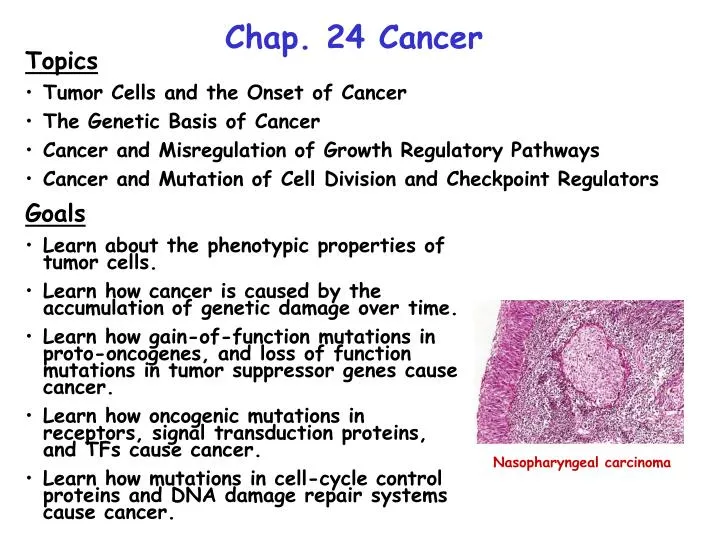
Chap. 24 Cancer. Topics Tumor Cells and the Onset of Cancer The Genetic Basis of Cancer Cancer and Misregulation of Growth Regulatory Pathways Cancer and Mutation of Cell Division and Checkpoint Regulators. Goals Learn about the phenotypic properties of tumor cells.

E N D
Chap. 24 Cancer • Topics • Tumor Cells and the Onset of Cancer • The Genetic Basis of Cancer • Cancer and Misregulation of Growth Regulatory Pathways • Cancer and Mutation of Cell Division and Checkpoint Regulators • Goals • Learn about the phenotypic properties of tumor cells. • Learn how cancer is caused by the accumulation of genetic damage over time. • Learn how gain-of-function mutations in proto-oncogenes, and loss of function mutations in tumor suppressor genes cause cancer. • Learn how oncogenic mutations in receptors, signal transduction proteins, and TFs cause cancer. • Learn how mutations in cell-cycle control proteins and DNA damage repair systems cause cancer. Nasopharyngeal carcinoma
Intro to Cancer Cancer is caused by the failure of genetic mechanisms that control the growth and proliferation of cells. In most cases, cumulative damage to multiple genes (the "multi-hit" model) via physical and chemical agents, replication errors, etc. contribute to oncogenesis. However, a person's inherited genetic background also may strongly contribute. In cancer, a single transformed cell grows to become a primary tumor, accumulates more mutations and becomes more aggressive, then metastasizes to another tissue and forms a secondary tumor. Metastasis usually is nonrandom in that tissues that produce growth factors and angiogenic factors are most susceptible to invasion. The difference between a benign tumor and a malignant one mostly involves the latter's ability to invade and metastasize to other tissues. Tumors are classified according to the embryonic origin of the tissue from which they originate. The term carcinoma is used to denote cancers of endodermal (e.g., gut epithelia cancers) or ectodermal (e.g., skin, neural epithelia) origin. Cancers of mesodermal origin (e.g., muscle, blood cells) are called sarcomas. Carcinomas make up >90% of malignant tumors.
Common Properties of Cancer Cells Cancer cells typically exhibit the alterations listed in Fig. 24.1. The most aggressive cancer cells display all of these features. Alterations are caused by mutations that affect growth factor receptors and signal transduction genes, cell cycle regulatory genes, DNA repair genes, or genes controlling apoptosis. Depending on whether the affected gene normally stimulates or inhibits proliferation, the mutated gene is called an oncogene or a tumor-suppressor gene. Cancers typically originate in dividing cells, such as precursor (progenitor) stem cells that give rise to differentiated tissues in the adult. Most cancers originate in somatic cells and not germ-line cells.
Tumor and Tumor Cell Morphology Very often (particularly in early stages) the diagnosis of cancer involves a histological examination of the affected tissue (Fig. 24.2). Microscopically, cancer cells show abnormal location and morphology, loss of structural features characteristic of a differentiated cell, and an abnormally large nucleus and prominent nucleoli. The cytoskeleton typically also is abnormal. Karyotyping may show gross chromosomal abnormalities. Advanced tumors such as the metastatic lung tumor of the liver shown in Fig. 25.2 are obvious. Such large tumors, in fact tumors larger than 2 mm in diameter, develop their own blood supply. To do this, cancerous cells secrete growth and angiogenic factors such as basic fibroblast growth factor (b-FGF), transforming growth factor a (TGFa), and vascular endothelial growth factor (VEGF).
Metastasis Most malignant tumor cells eventually acquire the ability to metastasize. To do so they must degrade the basement membranes of connective tissue underlying epithelial cells and surrounding the endothelial cells of blood vessels (Fig. 24.3). This can be accomplished by secretion of plasminogen activator, which activates the blood protease, plasmin. Malignant cells form structures called invadopodia which contain protein components needed for crossing basement membranes.
Cancer Stem Cells As mentioned above, cancers are thought to typically originate in dividing cells, such as precursor (progenitor) stem cells that give rise to differentiated tissues in the adult. These cells already possess one of the key properties needed for malignancy--the ability to continue to divide indefinitely. Recent research has shown that tumors do not contain a single type of transformed cell, although all are thought to come from a single original cell. Instead, tumors contain dangerous cancer stem cells that divide to produce a copy of themselves, and another cell of more limited replicative potential. Unfortunately, many cancer therapies that shrink tumors kill the more differentiated cells, leaving the cancer stem cells alive and capable of seeding additional tumors.
Multi-hit Model for Cancer Induction (I) The "multi-hit" model for cancer induction theorizes that metastatic tumor cells evolve from an original transformed cell via the accumulation of multiple mutations that increase its survivability and invasion potential. The multiple mutation theory is supported by the fact that the incidence of contracting most cancers increases steadily with age (Fig. 24.6).
Multi-hit Model for Cancer Induction (II) Studies with transgenic mice also support the multi-hit model for cancer induction. As shown in Fig. 24.7, the combined expression of the rasV12 oncogene and over-expression of the mycproto-oncogene causes a higher frequency of tumors in mice than when either gene is expressed alone. In the experiment shown, both genes were placed under the control of the strong breast-specific promoter derived from mouse mammary tumor virus. These genes are turned on in female mice at ~25 days of age when they are impregnated. The fact that tumor incidence increases with age for all 3 types of mice suggests that other accumulated mutations are contributing to cancer induction. myc encodes a TF that is induced by MAP kinase signaling. myc induces the expression of genes needed for the G1 S transition of the cell cycle.
Multi-hit Model for Cancer Induction (III) Compelling evidence for the multi-hit model comes from the study of the progression of lesions in the development of human colon cancer (Fig. 24.8). Cancers begin as a benign polyp that grows and becomes a benign adenoma. Later the adenoma turns into a carcinoma. Genetic analysis of cells from each stage shows that loss-of-function mutations in the APCtumor-suppressor gene (APC, adenomatous polyposis coli) occur in all polyps. APC- and rasD mutations occur in the benign adenoma stage. Later, loss-of-function of the p53 tumor-suppressor gene results in a malignant carcinoma with metastatic properties.
DNA Microarray Analysis of Gene Expression in Cancer Cells Many cancers have a different malignant potential despite being histologically indistinguishable. Furthermore, the exact combination of mutations and even genes altered in a given type of cancer can differ from one individual to another. Microarray analysis is now being used to study gene expression patterns (“signatures”) in cancers (Fig. 24.10). In the future, this will allow physicians to select the most appropriate treatment regimens (e.g., combination of drugs) to eliminate or control the cancer.
Genes Commonly Mutated in Cancer Genes that control cell growth and proliferation are commonly mutated in cancers (Fig. 24.11). Gain-of-function mutations that increase the activity of proto-oncogenes such as growth-promoting signaling molecules (I), receptors (II), intracellular signal transduction pathways (III), or TFs (IV) are associated with cancers. The resulting mutated genes are referred to as oncogenes. Loss-of-function mutations in tumor-suppressor genes such as cell cycle control proteins (V), DNA repair proteins (e.g., caretaker genes, VI), or anti-proliferative factor receptors such as the TGFß receptor can cause cancer. Lastly, gain-of-function mutations in anti-apoptotic genes and loss-of-function mutations in pro-apoptotic genes are associated with cancer (VII). Although relatively rare, human cancers can be caused by retroviruses that carry dominant viral oncogenes. More commonly, the insertion of a strong retroviral promoter upstream of a proto-oncogene can switch it on leading to cancer.
Generation of Oncogenes Oncogenes are generated from proto-oncogenes through gain-of-function mutations. Such mutations may create a constitutively active protein product (oncoprotein). Chromosomal translocations can create unregulated chimeric oncoproteins or place a proto-oncogene under the control of an uncontrolled promoter. DNA containing a proto-oncogene can be amplified, leading to over-expression of the transforming gene product. The latter is illustrated in Fig. 24.12a for the N-myc oncogene. Staining shows that myc DNA on one copy of chromosome 4 isolated from a human neuroblastoma cell is greatly amplified. Amplified DNA also can occur in mini-chromosomes known as minute chromosomes. Gene amplification can be assayed by microarray-type techniques that measure gene copy number changes. Gene amplifications are thought to arise from DNA replication errors.
Inherited Mutations in Tumor-suppressor Genes About 10% of human cancers have a hereditary basis. In most cases, the patient inherits one non-functional copy of a tumor-suppressor gene. Cancer is induced after the second functional copy of the gene is inactivated by mutation (loss of heterozygosity). Mutations in additional genes typically also are required. The induction of hereditary vs sporadicretinoblastoma, which involves the RB gene, is compared in Fig. 24.13. The hereditary form of this cancer usually appears in childhood in both eyes. The sporadic form (which requires two somatic mutations) occurs later in life, and in only one eye. Similarly, loss-of-function mutations in one copy of the APCand BRCA genes predispose individuals to develop colon and breast cancer, respectively. Women with one mutant allele of BRCA have a 60% probability of developing breast cancer by age 50. BRCA encodes a protein that is important in DNA repair.
Other Loss of Heterozygosity Processes Two other loss of heterozygosity mechanisms for cancer induction in inherited cancers are illustrated in Fig. 24.14. First, nondisjunction events during mitosis in a somatic cell can result in one of the daughter cells receiving two chromosomes carrying the mutant allele. Second relatively rare mitotic recombination events can result in a daughter cell receiving two copies of the mutant allele. In both cases, cancer then can be induced in the cell homozygous for the mutant allele. Cancer also can be caused by deletion of the functional copy of a tumor suppressor gene.
Oncogenic Mutations in Her Receptors Although mutations that activate signal molecules are quite rare, oncogenic mutations that constitutively turn on receptors are common. For example, mutations that cause dimerization of RTKs and turn on their intrinsic tyrosine kinase activity have been described (Fig. 24.17). For example, mutations that lead to dimerization of the Her2 receptor for an EGF-related molecule in mouse mammary gland cause breast cancer. In humans, over-expression of the Her2 receptor also has been detected in some breast cancers. This illustrates how the activation of a protein or its over-expression can cause cancer.
Viral Activators of Receptors Viral oncoproteins have been discovered that turn on cell division and proliferation by mimicking receptor ligands and binding to growth factor receptors. In mice, the spleen focus-forming virus synthesizes a viral protein, gp55, that binds to erythropoietin (Epo) receptors in erythroid progenitor cells causing them to dimerize and activate associated JAK kinases (Fig. 24.18). This causes excessive RBC production and leads to erythroleukemia. Human papillomavirus (HPV), which causes genital warts and cervical cancer, produces a protein that triggers growth of cells via activation of the platelet derived growth factor (PDGF) receptor. A vaccine against HPV recently was approved for use.
Oncogenic Signal Transduction Proteins The first non-viral oncoprotein discovered was RasD. RasD mutations result from amino acid substitutions that inhibit the GTPase activity of Ras or its interactions with GAP proteins. In both cases, RasD is locked in an active form. Constitutively activated RasD proteins occur in many bladder, colon, mammary, skin, and lung cancers, and in leukemias. Cancer-causing mutations also have been found in Ras-associated Raf kinase and GAP genes. Actually, the first oncoprotein discovered was the v-Src viral tyrosine kinase encoded by Raus sarcoma virus (Fig. 24.19). v-Src is derived from c-Src, a member of a family of cytosolic tyrosine kinases. Through the accumulation of mutations, the v-Src species became constitutively active. Cells undergo cancerous transformation after infection by the virus.
Chromosomal Translocations in Cancer Chronic myelogenous leukemia (CML) commonly is caused by a chromosomal translocation between chromosomes 9 and 22 that generates the Philadelphia chromosome (Fig. 24.20a). The translocation creates a hybrid kinase (the BCR-ABL fusion protein) that phosphorylates and activates many signal transduction proteins leading to cell proliferation. The drug imatinib (Gleevec) inhibits the kinase and is effective in controlling CML. In Burkitt's lymphoma, a translocation between chromosomes 8 and 14 places the c-myc gene under the control of the enhancer for the antibody heavy chain gene (CH) (Fig. 24.22). This results in high expression of c-myc and excessive cell division leading to cancer.
Loss-of-function Mutations in the TGFß Signaling Pathway TGFß is an anti-proliferative factor that signals via the Smad signal transduction pathway (Fig. 24.23). Loss-of-function mutations in TGFß receptors and Smads result in decreased expression of genes that limit cell proliferation. For example, the p15 gene is a tumor-suppressor gene that encodes an inhibitor of G phase cyclin-CDKs. p15 thus is important for arrest of cells in G1. The PAI-1 gene encodes plasminogen activator inhibitor, which helps prevent degradation of the extracellular matrix. The loss of the expression of these genes and other TGFß-controlled genes contributes to cell transformation.
Mutations that De-regulate the G1 to S-phase Transition Many cancers exhibit mutations in genes that affect the regulation of G1 S START control of the cell cycle. As discussed earlier, the Rb protein plays a major role in regulating passage through the restriction point by inhibiting the activity of the E2F TF (Fig. 24.24). Many cancer cells contain loss-of-function mutations in the Rb gene. Cancer cells also may fail to synthesize the p16 protein. p16 inhibits G1 cyclin-CDKs (cyclin D-CDK4) which phosphorylate and inactivate Rb. Finally, tumors of antibody-producing B lymphocytes have been isolated which over-express cyclin D due to a chromosomal translocation that places the cyclin D gene under the control of an antibody gene enhancer.