Download

1 / 46
560 likes | 957 Views
Cardiac Biomarkers:. History. 1950’s : Clinical reports that transaminases released from dying myocytes could be detected via laboratory testing, aiding in the diagnosis of myocardial infarction 1

E N D
History • 1950’s: Clinical reports that transaminases released from dying myocytes could be detected via laboratory testing, aiding in the diagnosis of myocardial infarction1 • The race to define clinical markers to aid in the diagnosis, prognosis, and risk stratification of patients with potential cardiovascular disease begins 1Circulation 108:250-252
History • Initial serum markers included AST, LDH, total CK and α-hydroxybutyrate • These enzymes are all released in varying amounts by dying myocytes • Lack of sensitivity and specificity for cardiac muscle necrosis fuels continued research
History: CK and Isoenzymes • CK known to be released during muscle necrosis (including cardiac) • Quantitative assays were cumbersome and difficult to perform • Total CK designed as a fast, reproducible spectrophotometric assay in the late 1960’s
History: CK and Isoenzymes • CK isoenzymes are subsequently described • MM, MB and BB fractions • 1970’s: MB fraction noted to be elevated in and highly specific for acute MI1 1 Clinical Chemistry 50(11): 2205-2213
History: CK and Isoenzymes • CKMB now measured via a highly sensitive monoclonal antibody assay • It was felt for a time that quantitative CKMB determination could be used to enzymatically measure the size of an infarct • This has been complicated by release of additional enzymes during reperfusion
History: CK and Isoenzymes • As CK-MB assays become more sensitive, researchers come to the paradoxical realization that it too is not totally cardiac specific • The MB fraction is determined to be expressed in skeletal muscle, particularly during the process of muscle regeneration • The search for cardiac specificity continues… Clinical Chemistry 50(11): 2205-2213
History • Research turns towards isolation of and development of assays for sarcomeric proteins • Myosin light chains were originally isolated and then subsequently abandoned because of specificity issues
History: Troponin • Troponin I first described as a biomarker specific for AMI in 19871; Troponin T in 19892 • Now the biochemical “gold standard” for the diagnosis of acute myocardial infarction via consensus of ESC/ACC 1 Am Heart J 113: 1333-44 2 J Mol Cell Cardiol 21: 1349-53
History • This work encourages development of other clinical assays for diagnosis and prognosis of a wide spectrum of cardiac diseases • Notable examples: • BNP (FDA approved in November 2000 for diagnosis of CHF) • C-reactive protein
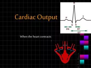
What is Myocardial Infarction? • Myocardial ischemia results from the reduction of coronary blood flow to an extent that leads to insufficiency of oxygen supply to myocardial tissue • When this ischemia is prolonged & irreversible, myocardial cell death & necrosis occurs ---this is defined as: myocardial infarction
Biochemical Changes in Acute Myocardial Infarction(mechanism of release of myocardial markers) ischemia to myocardial muscles (with low O2 supply) anaerobic glycolysis increased accumulation of Lactate decrease in pH activate lysosomal enzymes disintegration of myocardial proteins cell death & necrosis ECG changes clinical manifestations (chest pain) release of intracellular contents to blood BIOCHEMICAL MARKERS
Diagnosis of Myocardial Infarction SHOULD depend on THREE items (as recommended by WHO) 1- Clinical Manifestations 2- ECG 3- Biochemical Markers
Markers of Cardiac Necrosis • Cardiac biomarkers an integral part of the most recent joint ACC/ESC consensus statement on the definition of acute or recent MI:
“Perfect” Cardiac Marker • Early appearance • Accurate, specific, precise • Readily available, fast results • Cost-effective
Markers of Cardiac Necrosis Typical rise and gradual fall (troponin) or more rapid rise and fall (CK-MB) of biochemical markers of myocardial necrosis with at least (1) of the following: • Ischemic symptoms • Development of pathologic Q waves • ST segment elevation or depression • Coronary artery intervention
Troponins • Troponin T (cTnT) and troponin I (cTnI) control the calcium-mediated interaction of actin and myosin • cTnI completely specific for the heart • cTnT released in small amounts by skeletal muscles, though clinical assays do not detect skeletal TnT
Troponins • 4-6 hours to rise post-infarct, similar to CKMB • 6-9 hours to detect pathologic elevations in all patients with infarct • Elevated levels can persist in blood for weeks; the cardiac specificity of troponins thus make them the ideal marker for retrospective diagnosis of infarction
CK-MB • High specificity for cardiac tissue • The preferred marker for cardiac injury for many years • Begins to rise 4-6 hours after infarction but can take up to 12 hours to become elevated in all patients with infarction • Elevations return to baseline within 36-48 hours, in contrast to troponins • CK-MB is the marker of choice for diagnosis of reinfarction after CABG because of rapid washout
CK-MB: Shortcomings • Concomitant skeletal muscle damage can confuse the issue of diagnosis: • CPR and defibrillation • Cardiac and non-cardiac procedures • Blunt chest trauma • Cocaine abuse
CK:CK-MB Ratio • Proposed to improve specificity for use in diagnosis of AMI • Ratios 2.5-5 have been proposed • Significantly reduces sensitivity in patients with both skeletal muscle and cardiac injury • Also known to be misleading in the setting of hypothyroidism, renal failure, and chronic skeletal muscle diseases
Myoglobin • Heme protein rapidly released from damaged muscle • Elevations can be seen as early as one hour post-infarct • Much less cardiac specific; meant to be used as a marker protein for early diagnosis in conjunction with troponins
Natriuretic Peptides • Present in two forms, atrial (ANP) and brain (BNP) • Both ANP and BNP have diuretic, natriuretic and hypotensive effects • Both inhibit the renin-angiotensin system and renal sympathetic activity • BNP is released from the cardiac ventricles in response to volume expansion and wall stress
BNP Assay • Approved by the FDA for diagnosis of cardiac causes of dysnpea • Currently measured via a rapid, bedside immunofluorescence assay taking 10 minutes • Especially useful in ruling out heart failure as a cause of dyspnea given its excellent negative predictive value
BNP • Came to market in 2000 based on data from many studies, primarily the Breathing Not Properly (BNP) study • Prospective study of 1586 patients presenting to the ER with acute dyspnea • The predictive value of BNP much superior to previous standards including radiographic, clinical exam, or Framingham Criteria
BNP • BNP has also shown utility as a prognostic marker in acute coronary syndrome • It is associated with increased risk of death at 10 months as concentration at 40 hours post-infarct increased • Also associated with increased risk for new or recurrent MI
Prognostic Markers and Markers of Risk Stratification • C-reactive protein • Myeloperoxidase • Homocysteine • Glomerular filtration rate
C-Reactive Protein • Multiple roles in cardiovascular disease have been examined • Screening for cardiovascular risk in otherwise “healthy” men and women • Predictive value of CRP levels for disease severity in pre-existing CAD • Prognostic value in ACS
C-Reactive Protein • Pentameric structure consisting of five 23-kDa identical subunits • Produced primarily in hepatocytes • Plasma levels can increase rapidly to 1000x baseline levels in response to acute inflammation • “Positive acute phase reactant”
C-Reactive Protein • Binds to multiple ligands, including many found in bacterial cell walls • Once ligand-bound, CRP can: • Activate the classical compliment pathway • Stimulate phagocytosis • Bind to immunoglobulin receptors
C-Reactive Protein:Risk Factor or Risk Marker? • CRP previously known to be a marker of high risk in cardiovascular disease • More recent data may implicate CRP as an actual mediator of atherogenesis • Multiple hypotheses for the mechanism of CRP-mediated atherogenesis: • Endothelial dysfunction via ↑ NO synthesis • ↑LDL deposition in plaque by CRP-stimulated macrophages
CRP and CV Risk • Elevated levels predictive of: • Long-term risk of first MI • Ischemic stroke • All-cause mortality
Myeloperoxidase • Released by activated leukocytes at elevated levels in vulnerable plaques • Predicts cardiac risk independently of other markers of inflammation • May be useful in triage of ACS (levels elevate in the 1st two hours) • Also identifies patients at increased risk of CV event in the 6 months following a negative troponin NEJM 349: 1595-1604
Homocysteine • Intermediary amino acid formed by the conversion of methionine to cysteine • Moderate hyperhomocysteinemia occurs in 5-7% of the population • Recognized as an independent risk factor for the development of atherosclerotic vascular disease and venous thrombosis • Can result from genetic defects, drugs, vitamin deficiencies, or smoking
Homocysteine • Homocysteine implicated directly in vascular injury including: • Intimal thickening • Disruption of elastic lamina • Smooth muscle hypertrophy • Platelet aggregation • Vascular injury induced by leukocyte recruitment, foam cell formation, and inhibition of NO synthesis
Homocysteine • Elevated levels appear to be an independent risk factor, though less important than the classic CV risk factors • Screening recommended in patients with premature CV disease (or unexplained DVT) and absence of other risk factors • Treatment includes supplementation with folate, B6 and B12
Glomerular Filtration Rate • The relationship between chronic kidney disease and cardiovascular risk is longstanding • Is this the result of multiple comorbid conditions (such as diabetes and hypertension), or is there an independent relationship?
Glomerular Filtration Rate • Recent studies have sought to identify whether creatinine clearance itself is inversely related to increased cardiovascular risk, independent of comorbid conditions
Glomerular Filtration Rate • Go, et al performed a cohort analysis of 1.12 million adults in California with CKD that were not yet dialysis-dependent • Their hypothesis was that GFR was an independent predictor of cardiovascular morbidity and mortality • They noted a strong independent association between the two NEJM 351: 1296-1305
Glomerular Filtration Rate • Reduced GFR has been associated with: • Increased inflammatory factors • Abnormal lipoprotein levels • Elevated plasma homocysteine • Anemia • Arterial stiffness • Endothelial dysfunction