Download
1 / 14
150 likes | 724 Views
Gram-Positive Bacilli Part Two. MLAB 2434: Microbiology Keri Brophy -Martinez. Rods. Erysipelothrix Lactobacillus Gardnerella vaginalis. Erysipelothrix rhusiopathiae: General characteristics. Gram positive, non–spore-forming, pleomorphic rods (can produce long filaments)

E N D
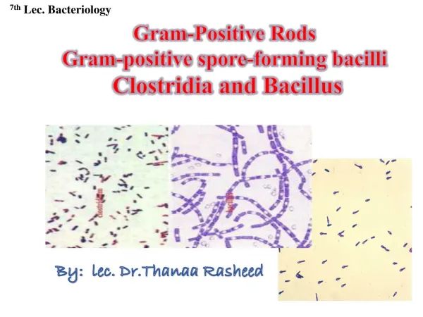
Gram-Positive BacilliPart Two MLAB 2434: Microbiology Keri Brophy-Martinez
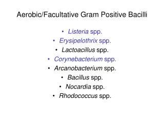
Rods Erysipelothrix Lactobacillus Gardnerella vaginalis
Erysipelothrix rhusiopathiae:General characteristics • Gram positive, non–spore-forming, pleomorphic rods (can produce long filaments) • Distributed in nature • Can cause disease in animals (swine, turkey, sheep); swine is the main reservoir • Humans acquire the infection through occupational exposure, such as cuts & scratches (fish handlers, animal products)
Erysipelothrix rhusiopathiae:Clinical Infections • Erysipeloid • Self-limiting localized infection at the site of inoculation • Produces painful swelling, usually on the hands or fingers • Heals within 3 to 4 weeks • Treat with penicillin, cephalosporin, erythromycin • Endocarditis • May occur in those who have had valve replacements • Disseminated infections may occur, but rarely
Laboratory Diagnosis: Erysipelothrix rhusiopathiae • Colony Morphology • CO2 is required • Grows on blood or chocolate agar • Colonies may appear gray or translucent, pinpoint • Alpha hemolysis or nonhemolytic
Laboratory Diagnosis:Erysipelothrix rhusiopathiae • Microscopic Morphology • Pleomorphic, • Gram-positive thin rods that may form long filaments or short rods • Arranged singly, in short chains, or in a V shape
Laboratory Diagnosis: Erysipelothrix rhusiopathiae • Identification • Catalase, nitrate, urease negative • Nonmotile • Production of H2S on TSI • Test tube brush growth in semisolid motility media
Characteristics of Corynebacterium, Listeria, and Erysipelothrix
Lactobacillus • Widely distributed in nature • Normal flora of mouth, GI tract and female genital tract • Treat with pencillin plus an aminoglycoside • resistant to vancomycin (helps in diagnosis) • Clinical Infections • Bacterial vaginosis • Bacteremia, endocarditis, meningitis (rare)
Lactobacillus • Microscopic Morphology • Long, slender gram positive pleomorphic bacilli • Non-spore forming • Colony Morphology • SBA • Pinpoint • α- hemolytic or gamma colonies • Lab Diagnosis • Catalase negative
Gardnerella vaginalis • Member of the normal flora of the female genital tract • Associated with bacterial vaginosis • Foul odor • Vaginal pH > 4.5
Laboratory Diagnosis:Gardnerellavaginalis • Wet Prep • Look for clue cells • Large epithelials with various bacterial types on edges • Gram stain • Small, thin • Gram variable rods
Laboratory Diagnosis:Gardnerellavaginalis • Cultural Characteristics • Growth on BAP, CA • No growth on MAC • Human blood bilayertween “V” agar • Beta-hemolytic • Requires a CO2 environment • Catalase negative
References • Engelkirk, P. G., & Duben-Engelkirk, J. (2008). Laboratory Diagnosis of Infectious Diseases: Essentials of Diagnostic Microbiology . Baltimore, MD: Lippincott Williams & Willkins. • http://en.wikipedia.org/wiki/Lactobacillus • http://www.thefullwiki.org/Corynebacterium_diphtheriae • http://quizlet.com/10262287/print/ • Kiser, K. M., Payne, W. C., & Taff, T. (2011). Clinical Laboratory Microbiology: A Practical Approach . Upper Saddle River, NJ: Pearson Education, Inc. • Mahon, C. R., Lehman, D. C., & Manuselis, G. (2011). Textbook of Diagnostic Microbiology (4th ed.). Maryland Heights, MO: Saunders.