Download
1 / 44
450 likes | 608 Views
SECONDARY HYPERTENSION. DEFINITION. Essential, primary, or idiopathic hypertension is defined as high BP in which secondary causes or mendelian (monogenic) forms are not present High BP – repeatedly measured BP exceeding 140/90 mmHg, i.e. a systolic BP above 140 and/or diastolic BP above 90.

E N D
DEFINITION • Essential, primary, or idiopathic hypertension is defined as high BP in which secondary causes or mendelian (monogenic) forms are not present • High BP – repeatedly measured BP exceeding 140/90 mmHg, i.e. a systolic BP above 140 and/or diastolic BP above 90
Aetiology of Hypertension • Primary – 90-95% of cases – also termed “essential” of “idiopathic” • Secondary – about 5% of cases • Renal or renovascular disease • Endocrine disease • Phaeochomocytoma • Cushings syndrome • Conn’s syndrome • Acromegaly and hypothyroidism • Coarctation of the aorta • Iatrogenic • Hormonal / oral contraceptive • NSAIDs
Aetiology of Hypertension • Primary – 90-95% of cases – also termed “essential” of “idiopathic” • Secondary – about 5% of cases • Renal parenchymal (2-5%) or renovascular disease • Endocrine disease • Phaeochomocytoma • Cushing syndrome • Conn syndrome • Acromegaly and hypothyroidism • Coarctation of the aorta • Iatrogenic • Hormonal / oral contraceptive • NSAIDs
Renal parenchymal disease • Acute and chronic glomerulonephritis • Polycystic kidney disease • Diabetic nephropathy • Pyelonephritis • Obstructive uropathy • Neoplasms • Renal trauma • Radiation nephritis
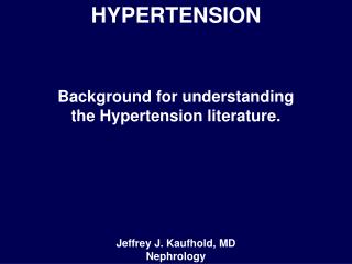
Renal parenchymal disease CIN – chronic interstitial nephritis; APKD – adult-onset polycystic kidney disease; MCN - minimal change nephropathy; MGN – membranous glomerulonephritis; DN – diabetic nephropathy; MPGN – membranoproliferative glomerulonephritis; FSGN – focal segmental glomerulonephritis
Candidate pathophysiologic mechanisms related to hypertension in parenchymal renal disease
Hypertension in parenchymal renal disease: major target organ manifestations
Hypertension in parenchymal renal disease:CONCLUSIONS • Hypertension may result from renal disease that reduces functioning nephrons; • Evidence shows a clear relationship between high blood pressure and end-stage renal disease; • BP should be controlled to 130/85 mmHg (125/75 mmHg in patients with proteinuria in excess of 1g/24 h)
Aetiology of Hypertension • Primary – 90-95% of cases – also termed “essential” of “idiopathic” • Secondary – about 5% of cases • Renal parenchymal or renovascular disease (0.3-3%) • Endocrine disease • Phaeochomocytoma • Cushings syndrome • Conn’s syndrome • Acromegaly and hypothyroidism • Coarctation of the aorta • Iatrogenic • Hormonal / oral contraceptive • NSAIDs
RENAL ARTERY STENOSIS(RAS) • Atherosclerotic RAS (>90% of cases): involves the ostium and the proximal portion of the main renal artery with plaque extending into the perirenal aorta • Fibromuscular dysplasia (10% of cases): typically seen in young and middle-aged females. As opposed to atherosclerotic RAS, fibromuscular dysplasia typically affects the distal two thirds of the main renal artery
RENAL ARTERY STENOSIS:screening and diagnostic studies • Renal duplex sonography • Magnetic resonance angiography • Renal artery arteriography • Captopril renography
RENAL ARTERY STENOSIS:renal duplex sonography Stenoses over 60%: • Peak systolic velocity (PSV) >150-180 cm/sec • Renal-aortic ratio >3.5 Prognostic value: • Resistance index (RI): RI=(1-EDV)/PSVx100; if RI>80 no benefit after revascularization
RENAL ARTERY STENOSIS:MR angiography Strong sides: • Provides images of the renal arteries, 3D-reconstruction, plaque characterization and hemodynamic information • Gadolinium (contrast agent): non-nephrotoxic Weak sides: high cost and limited availability
RENAL ARTERY EVALUATION:contrast angiography (the “gold” standard) Fibromuscular dysplasia: “string of beads” appearance Atherosclerotic RAS with poststenotic dilatation
RENAL ARTERY STENOSIS:treatment • BP control • Antiplatelet, lipid-lowering therapy, and beta-blockers, if appropriate • No ACE-inhibitors in severe RAS !
RENAL ARTERY STENOSIS:treatment Percutaneous or surgical revascularization, if: ● Resistant or poorly controlled hypertension and unilateral or bilateral renal artery stenosis ● Renal artery stenosis and recurrent flash pulmonary edema for which there is no readily explainable cause ● Chronic renal failure and bilateral renal artery stenosis or renal artery stenosis to asolitary functioning kidney ● Sonographic renal longitudinal length >7cm
Aetiology of Hypertension • Primary – 90-95% of cases – also termed “essential” of “idiopathic” • Secondary – about 5% of cases • Renal or renovascular disease • Endocrine disease • Phaeochomocytoma (0.1-0.6 %) • Cushings syndrome • Conn’s syndrome • Acromegaly and hypothyroidism • Coarctation of the aorta • Iatrogenic • Hormonal / oral contraceptive • NSAIDs
PHEOCHROMOCYTOMA“frequently searched for, but rarely found” • About 90 % of pheochromocytomas are located within the adrenal glands; • 10% are bilateral; • 10% are malignant; • 10% are extra-adrenal; • Extra-adrenal pheochromocytomas develop in paraganglion chromaffin tissue of the sympathetic nervous system; of them, 40% are not diagnosed, 5% are multiple; • overall, nearly 98% of pheochromocytomas are found in the abdomen
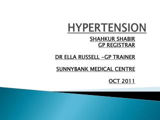
PHEOCHROMOCYTOMA“the great mimic” Frequency of signs and symptoms (%) of pheochromocytoma
PHEOCHROMOCYTOMAdiagnostic techniques • Biochemical tests • High pressure liquid chromatography: • Plasma catecholamines: noradrenaline, adrenaline; • Free plasma fractionated metanephrines: normetanephrine, metanephrine; • Urinary catecholamines (24h) • Urinary fractionated metanephrines (24h) • Spectrophotometry: • Total metanephrines (24h urine); • Vanillylmandelic acid(24h urine)
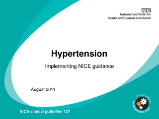
PHEOCHROMOCYTOMA Sensitivity and specifity of biochemical tests for diagnosis of pheochromocytoma
PHEOCHROMOCYTOMAimaging techniques • Duplex sonography; • Magnetic resonance imaging (MRI); • Computed romography (CT); • 123I – meta-iodo-benzyl-guanidine scanning (123I-MIBG)
PHEOCHROMOCYTOMA Sonography : • Sonographic appearances are those of a well-defined homogeneous hypoechoic mass in approximately 50 pet cent of patients. • However the mass may be complex or even cystic (16 pet cent) and hyperechoic to the renal parenchyma (approximately 20 pet cent).
PHEOCHROMOCYTOMA MRI (coronal and sagittal sections): • Magnetic resonance (MR) imaging is equally sensitive to CT and lends itself to in vivo tissue characterization, which is not possible with CT; • MR imaging is nearly 100% sensitive and around 70% specific. • Preferred for the localisation of extra-adrenal tumours or tumours during pregnancy, in children, or in patients with allergies to contrast
PHEOCHROMOCYTOMA CT: • accurately detects tumors larger than 1.0 cm and has a localization precision of approximately 98%, although it is only 70% specific; • since CT scanning and MRI have similar sensitivities (90–100%) and specificities (70–80%), MRI is the preferred procedure
PHEOCHROMOCYTOMA 123I-MIBG scanning: • increased specificity (95–100%), as compared with CT or MRI; • provides both anatomic and functional characterization; • Relevant in patients with multiple, extra-adrenal, malignant (metastatic) tumors
PHEOCHROMOCYTOMA: laparoscopic removal Preoperative Management (10-14 days) • Purpose:to prevent catecholamine induced, serious, and potentially life-threatening complications during surgery, including hypertensive crises, cardiac arrhythmias, pulmonary oedema, and cardiac ischemia; • BP should be reduced to below 160/90 mm Hg for at least 24h; • orthostatic hypotension should be present, but blood pressure in the upright position should not fall below 80/45 mm Hg; • there should be no more than one ventricular extrasystole every 5 min; • and the electrocardiogram should show no S-T segment changes and T-wave inversions for 1 week;
PHEOCHROMOCYTOMA: Management • Phenoxybenzamine, a long acting alpha-adrenergic blocker, is the mainstay of medical treatment to control BP. A total dose of 1 mg/kg is sufficient in most patients. • An alpha-blocker Doxazosin in increasing doses from 1 to 16 mg once a day. • A beta-adrenoceptor blocker (eg,propranolol 40 mg three times daily or atenolol 25–50 mg once daily) could be included after several days of alpha-adrenergic blockade. • Adequate salt and fluid intake lowers the risk of orthostatic hypotension.
PHEOCHROMOCYTOMA: Management • Should substantial rises in blood pressure still take place during surgery, these can be controlled by bolus or by continuous infusion of phentolamine, sodium nitroprusside, or a shortacting calcium antagonist (eg, nicardipine); • Tachyarrhythmias can be treated by infusion of a shortacting -adrenoceptor blocker (eg, esmolol).
PHEOCHROMOCYTOMA Sensitivity and specifity of biochemical tests for diagnosis of pheochromocytoma
Conn’s Syndrome (primary hyperaldosteronism) • Should be considered in any hypertensive pt with muscle weakness, polydipsia, andor hypokalemia; • 75% - adrenal adenoma; • 25% - adrenal hyperplasia • Rarely – adrenocortical cancer
Primary Hyperaldosteronism • Screening for hyperaldosteronism should include plasma aldosterone and plasma renin activity measured in morning samples • Plasma aldosterone:renin ratio: normally <20; diagnostic cut-off value >30; • Aldosterone excretion rate during salt loading, captopril, or spironolactone test (the captopril test may be less useful in blacks because of the high prevalence of low plasma renin activity) • Adrenal CT, MRI
Primary Hyperaldosteronism Should be differentiated from • Secondary hyperaldosteronism in patients with renal failure, CHF, essential hypertension • Monogenic forms of hypertension (pseudohyperaldosteronism): • Liddle’s syndrome (autosomal-dominant disorder, characterized by low-renin, low-aldosterone, low-potassium volume-expanded hypertension) • Gordon’s syndrome (autosomal-dominant disorder, characterized by low-renin, low-aldosterone, high-potassium volume-expanded hypertension)
Primary Hyperaldosteronism TREATMENT 1. Medical • Spironolactone, a competitive aldosterone antagonist • Amiloride, a potassium-sparing diuretic • Glucocorticoids (in glucocorticoid-remediable form) 2. Surgical, if appropriate
Aetiology of Hypertension • Primary – 90-95% of cases – also termed “essential” of “idiopathic” • Secondary – about 5% of cases • Renal or renovascular disease • Endocrine disease • Phaeochomocytoma • Cushing’s syndrome (0.1-0.6%) Conn’s syndrome • Acromegaly and hypothyroidism • Coarctation of the aorta • Iatrogenic • Hormonal / oral contraceptive • NSAIDs
Cushing’s Syndrome • Hypertension occurs in about 80% of patients; • Urinary free cortisol • If 24h UFC>100 µg/ml: measure plasma ACTH
Hypothyroidism • Both hypertension (particularly diastolic) and hypotension are common; Hyperthyroidism • Accompanied by systolic hypertension, especially in the elderly; Acromegaly • 25-50% exhibit elevated blood pressure