Download

1 / 9
100 likes | 225 Views
Synaptic Plasticity III. 4 April 2012. Housekeeping. NO LABS THIS WEEK! No lab 8 assignment due No class until next Wednesday Have a happy Easter! But a sad Good Friday! Problem Set 3 and Article Set 3 are available Problem Set 3 consists of questions about Article Set 3. Dendrites.

E N D
Synaptic Plasticity III 4 April 2012
Housekeeping • NO LABS THIS WEEK! • No lab 8 assignment due • No class until next Wednesday • Have a happy Easter! • But a sad Good Friday! • Problem Set 3 and Article Set 3 are available • Problem Set 3 consists of questions about Article Set 3
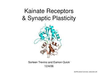
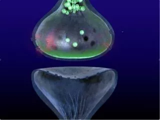
General Features of the Postsynaptic Specialization • Soma and dendrites are the predominant postsynaptic sites • Inhibitory synapsesare typically found on proximal dendrites and the soma • Excitatory synapses are typically found on spineson distal dendritic branches
Dendritic Spines: Structure and Function • Spines come in a variety of shapes and sizes • Functional significance of variety unclear • Most principle neurons display spines • Typically, GABAergic neurons do not display spines (aspiny) • Spines, therefore, represent the major postsynaptic site for excitatory input
Dendritic Spines:Structure and Function • Spines contain organelles: • Smooth ER—intracellular Ca2+ source • Polyribosomes and vesicles • Spines display the PSD • Approximately 10% of spine surface area • Precisely aligned with presynaptic active zone • Often associated with cell-cell adherens junctions
Dendritic Spines:Structure and Function • Each spine usually represents no more than a single synapse • Suggests synapse-specific signalling: Spines act as both segregators and integrators of synaptic signals • Spines are highly dynamic (seconds to minutes) through both activity-dependent and –independent mechanisms • Convincingly show experience-dependent morphological changes—memory?