Download
1 / 1
10 likes | 126 Views
F ACHHOCHSCHULE B RANDENBURG F ACHBEREICH I NFORMATIK UND M EDIEN. Automated segmentation of ophthalmological OCT images. Friedrich Müller , Reiner Creutzburg. Abstract:

E N D
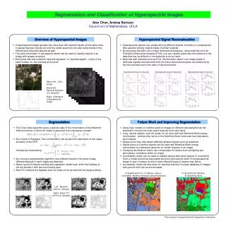
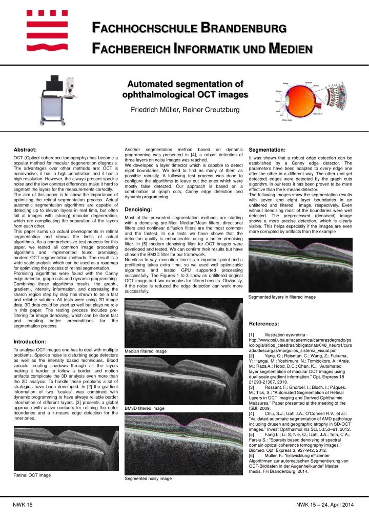
FACHHOCHSCHULE BRANDENBURG FACHBEREICH INFORMATIK UND MEDIEN Automated segmentation of ophthalmological OCT images Friedrich Müller, Reiner Creutzburg Abstract: OCT (Optical coherence tomography) has become a popular method for macular degeneration diagnosis. The advantages over other methods are: OCT is noninvasive, it has a high penetration and it has a high resolution. However, the always present speckle noise and the low contrast differences make it hard to segment the layers for the measurements correctly. The aim of this paper is to show the importance of optimizing the retinal segmentation process. Actual automatic segmentation algorithms are capable of detecting up to eleven layers in real time, but often fail at images with (strong) macular degeneration, which are complicating the separation of the layers from each other. This paper sums up actual developments in retinal segmentation and shows the limits of actual algorithms. As a comprehensive test process for this paper, we tested all common image processing algorithms and implemented found promising, modern OCT segmentation methods. The result is a wide scale analysis which can be used as a roadmap for optimizing the process of retinal segmentation. Promising algorithms were found with the Canny edge detector, graph cuts and dynamic programming. Combining these algorithms results, the graph-, gradient-, intensity information, and decreasing the search region step by step has shown to be a fast and reliable solution. All tests were using 2D image data, 3D data could be used as well but plays no role in this paper. The testing process includes pre-filtering for image denoising, which can be done fast and creating better preconditions for the segmentation process. Another segmentation method based on dynamic programming was presented in [4], a robust detection of three layers on noisy images was reached. We developed a layer detector which is capable to detect eight boundaries. We tried to find as many of them as possible robustly. A following test process was done to configure the algorithms to leave out the ones which were mostly false detected. Our approach is based on a combination of graph cuts, Canny edge detection and dynamic programming. Denoising: Most of the presented segmentation methods are starting with a denoising pre-filter. Median/Mean filters, directional filters and nonlinear diffusion filters are the most common and the fastest. In our tests we have shown that the detection quality is enhanceable using a better denoising filter. In [5] modern denoising filter for OCT images were developed and tested. We can confirm their results but have chosen the BM3D filter for our framework. Needless to say, execution time is an important point and a prefiltering takes extra time, so we used well optimizable algorithms and tested GPU supported processing successfully. The Figures 1 to 3 show an unfiltered original OCT image and two examples for filtered results. Obviously, if the noise is reduced the edge detection can work more successfully. Segmentation: It was shown that a robust edge detection can be established by a Canny edge detector. The parameters have been adapted to every edge one after the other in a different way. The other (not yet detected) edges were detected by the graph cuts algorithm, in our tests it has been proven to be more effective than the k-means detector. The following images show the segmentation results with seven and eight layer boundaries in an unfiltered and filtered image, respectively. Even without denoising most of the boundaries were well detected. The preprocessed (denoised) image shows a more precise detection, which is clearly visible. This helps especially if the images are even more corrupted by artifacts than the example Segmented layers in filtered image References: [1] Illustration eye/retina - http://www.psi.uba.ar/academica/carrerasdegrado/psicologia/sitios_catedras/obligatorias/048_neuro1/cursada/descargas/margulios_sistema_visual.pdf [2] Yang, Q.; Reisman, C.; Wang, Z.; Fukuma, Y; Hangai, M.; Yoshimura, N.; Tomidokoro, A.; Araie, M.; Raza A.; Hood, D.C.; Chan, K..: "Automatedlayersegmentationofmacular OCT imagesusing dual-scalegradientinformation." Opt. Express 18 21293-21307, 2010. [3]Rossant, F.; Ghorbel, I.; Bloch, I.; Pâques, M.; Tick, S.: "Automated Segmentation ofRetinalLayers in OCT Imaging andDerivedOphthalmicMeasures." Paper presentedatthemeetingofthe ISBI, 2009. [4]Chiu, S.J.; Izatt J.A.; O'Connell R.V.; et al.: “Validatedautomaticsegmentationof AMD pathologyincludingdrusenandgeographicatrophy in SD-OCT images.” InvestOphthalmolVisSci, 53:53–61, 2012. [5] Fang L.; Li, S; Nie, Q.; Izatt, J.A.; Toth, C.A.; Farsiu S.: "Sparsitybaseddenoisingofspectraldomainopticalcoherencetomographyimages." Biomed. Opt. Express 3, 927-942, 2012. [6] Müller, F.: “Entwicklung effizienter Algorithmen zur automatischen Segmentierung von OCT-Bilddaten in der Augenheilkunde“ Master thesis, FH Brandenburg, 2014. Introduction: To analyse OCT images one has to deal with multiple problems. Speckle noise is disturbing edge detectors as well as the intensity based techniques. Blood vessels creating shadows through all the layers making it harder to follow a border, and motion artifacts complicate the 3D analysis even more than the 2D analysis. To handle these problems a lot of strategies have been developed. In [2] the gradient information of two “scales” was combined with dynamic programming to have always reliable border information of different layers. [3] presents a global approach with active contours for refining the outer boundaries and a k-means edge detection for the inner ones. Median filtered image BM3D filtered image Retinal OCT image Segmentednoisyimage NWK 15 NWK 15 – 24. April 2014