Download
1 / 24
240 likes | 386 Views
Systemic Lupus Erythematosus. Justin A. Crocker AM Report 12/2/09. SLE. Autoimmune disease that affects multisystems 1.5 million cases of lupus Prevalence of 17 to 48 per 100,000 population Women > Men - 9:1 ratio 90% cases are women African Americans > Whites

E N D
Systemic Lupus Erythematosus Justin A. Crocker AM Report 12/2/09
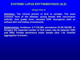
SLE • Autoimmune disease that affects multisystems • 1.5 million cases of lupus • Prevalence of 17 to 48 per 100,000 population • Women > Men - 9:1 ratio • 90% cases are women • African Americans > Whites • Onset usually between ages of 15 and 45 years, but • Can occur in childhood or later in life
Clinical Manifestations • For the purpose of identifying patients in clinical studies, a person has SLE if 4 or more of the 11 criteria are present, serially or simultaneously, during any interval of observation. (specificity 95%, sensitivity 75%) • It is important to remember that a patient may have SLE and not have 4 criteria.
Butterfly rash Discoid lupus Photosensitivity Oral ulcers Arthritis Serositis 7. Neurologic d/o 8. Hematologic d/o 9. Renal d/o 10.Immunologic: anti-DNA, anti-Sm, false pos STS 11.Anti-nuclear antibody Criteria
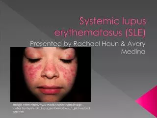
Cutaneous • Most common rash is photosensitive, raised erythematous malar rash. 55-85% develop at some point in disease • Discoid Lupus Erythematosus (DLE): 15-30% circular, scaly hyperpimented lesions with erythematous rim, atrophic center—can be disfiguring • Mouth/vaginal/nasal ulcers • Alopecia: may be diffuse or patchy. Occurs 50%
MSK • Polyarthritis, mild to disabling, occurs most frequently in hands, wrists, knees. Occurs 90% • Joint deformities occur in only 10% • Arthritis of SLE tends to be transitory • If single joint has persistent pain, consider osteonecrosis (prevalence increased in SLE over general population, especially if on steroids.) • Myositis with elevated CK and weakness rarely occurs
Serositis - Pulmonary • Pleuritis with or without effusion - if case is mild, tx: NSAIDS - if case is severe, tx: steroids • Life-threatening manifestations: interstitial inflammation which can lead to fibrosis and intra-alveolar hemorrhage. • Also pneumothorax and pulmonary HTN can occur
Serositis – Cardiac • Pericarditis: most common cardiac manifestation and usually responds to NSAIDs. • Myocarditis (rare) and fibrinous endocarditis (Libman-Sacks) may occur. Steroids plus treatment for CHF/arrhythmia or embolic events. • MI due to atherosclerosis can occur in <35 y/o
Neuro • Cranial or peripheral neuropathy occurs in 10-15%, it is probably secondary to vasculitis in small arteries supplying nerves. • Diffuse CNS dysfunction: memory and reasoning difficulty • Headache: if excruciating, often indicate acute flare • Seizures of any type • Psychosis: must distinguish from steroid-induced psychosis (occurs in 1st weeks of tx at doses ≥40mg prednisone and resolves after several days of reducing or stopping tx)
Cont. • TIA, Stroke: mostly increased among patients that are APLA positive • 50-fold increase in risk of vascular events in women under 45 compared to healthy women • Treatment for clotting event is long-term anticoagulation
Heme • Anemia: usually Normochromic, normocytic • Leukopenia: almost always consists of lymphopenia, not granulocytopenia • Thrombocytopenia
Renal • Nephritis: usually asymptomatic, so always check UA if patient has known or suspected SLE • Occurs early in course of disease-if not present w/in 1 yr, probably will not occur. • Histologic classification by renal biopsy is useful to plan therapy
Histologic Classifications • Class I is minimal mesangial glomerulonephritis which is histologically normal on light microscopy but with mesangial deposits on electron microscopy. • Class II is based on a finding of mesangial proliferative lupus nephritis. This form typically responds completely to treatment with corticosteroids. • Class III is focal proliferative nephritis and often successfully responds to treatment with high doses of corticosteroids. • Class IV is diffuse proliferative nephritis. This form is mainly treated with corticosteroids and immunosuppressant drugs. • Class V is membranous nephritis and is characterized by extreme edema and protein loss. • Class VI Glomerulosclerosis
Immunoglobulins • Anti-dsDNA IgG: very specific, may correlate with disease activity • Anti-Sm: specific, but only present in 25% of cases, does not correlate with activity • APLA: not specific. Used to identify patients at increased risk for clots, thrombocytopenia and fetal loss
ANA • ANA: positive in 95% of cases. Pretest probability affects interpretation. In PCP setting, 2% for SLE. In rheum: 30% • Low Positive (1:160 or lower): SLE likelihood <2% (<26% for rheumatologists) • High Positive (1:320 or higher): SLE likelihood: 2-17% (32-81% for rheumatologists) • SLE specific patterns: Rim and Homogenous
Additional work-up • Serum cr. and albumin • CBC w/ diff • U/A • ESR • Complement levels • Renal bx if warranted
Treatment • Treatment plans are based on patient age, sex, health, symptoms, and lifestyle • Goals of treatment are to: -prevent flares -treat flares when they occur -minimize organ damage and complications
Conservative management • For those w/out major organ involvement. • NSAIDs: to control pain, swelling, and fever • Caution w/ NSAIDS though. SLE pts are at increased risk for aseptic meningitis • Antimalarials: Generally to treat fatigue joint pain, skin rashes, and inflammation of the lungs • Commonly used: Hydroxycholorquine • Used alone or in combination with other drugs
Cont. • Corticosteroids (Mainstay of SLE treatment) • To rapidly suppress inflammation • Usually start with high-dose IV pulse and convert to PO steroids with goal of tapering and converting to something else. • Commonly used: prednisone, hydrocortisone, methylprednisolone, and dexamethasone
Immunosuppressives • Primarily for CNS/renal involvement • Mycophenolate mofetil (cellcept) • Azathioprine (imuran): requires several months to be effective, effective in smaller percentage of patients • MTX: for treatment of dermatitis and arthritis, not life-threatening disease • Cyclosporine: used in steroid-resistant SLE, risk of nephrotoxicity • Cyclophosphamide (cytoxan) Almost all trials performed on patients with nephritis