Download
1 / 72
720 likes | 850 Views
Interventions for Clients with Hematologic Problems. WHITE BLOOD CELL DISORDERS. White blood cells (WBCs), or leukocytes, provide protection from invading non-self cells and cancer cells in several ways.

E N D
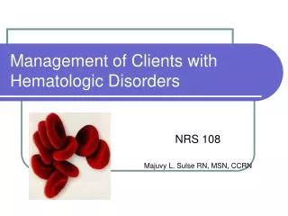
Interventions for Clients with Hematologic Problems WHITE BLOOD CELL DISORDERS
White blood cells (WBCs), or leukocytes, provide protection from invading non-self cells and cancer cells in several ways. • These protective functions depend on maintaining normal numbers and ratios of many specific mature, circulating leukocytes. • When any one type of WBC is present in either abnormally high or abnormally low amounts, hematopoietic function and immune function may be altered to some degree, placing clients at risk for specific complications
LEUKEMIA Overview • The leukemias are a group of malignant disorders involving abnormal overproduction of a specific WBC type, usually at an immature stage, in the bone marrow. • Leukemia may be acute, with a sudden onset and short duration, or chronic, with a slow onset and persistent symptoms over a period of years • Leukemias are categorized by the specific maturational pathway from which the abnormal cells arise. • Leukemias in which the abnormal cells arise from within the committed lymphoid maturational pathways are lymphocytic or lymphoblastic. • Leukemias in which the abnormal cells arise within the committed myeloid maturational pathways are myelocytic or myelogenous. • Several subtypes exist for each of these diseases, which are classified according to the degree of maturity of the abnormal cell and the specific cell type involved
Differentiating characteristics of the four types of leukemia
LEUKEMIA Pathophysiology • The basic problem in leukemia is a malignant transformation of the stem cells or early committed precursor leukocyte cells, causing an abnormal proliferation of a specific type of leukocyte. • The functionally and structurally abnormal immature leukocytes, produced in excessive quantities in the bone marrow, essentially shut down normal bone marrow production of erythrocytes, platelets, and other mature leukocytes. • This situation leads to anemia, thrombocytopenia, and leukopenia of the unaffected WBC types, even though the number of immature, abnormal WBCs in the circulation is greatly elevated. • Without treatment, the client usually dies of infection or hemorrhage. For clients with acute leukemias, these pathologic changes occur rapidly and, without intervention, progress quickly to death. Chronic leukemia may be present for many years before overt pathologic changes occur
LEUKEMIA Etiology • ionizing radiation • chemicals and drugs • marrow hypoplasia (slow functioning with less than the normal production rate of blood cells) • environmental interactions • genetic factors • viral factors • immunologic factors • the interaction of these factors
LEUKEMIA History • The nurse asks the client about: • risk factors and causative factors; • age; • occupation and hobbies; • previous illnesses and the medical history (may indicate exposure to ionizing radiation or medications that increase risk); • frequency and severity of infectious processes (such as colds, influenza, pneumonia, bronchitis, or unexplained episodes of fever) during the preceding 6 months; • any overt or hidden excessive bleeding episodes, such as the following: • a tendency to bruise easily • nosebleeds • increased menstrual flow • bleeding from the gums • rectal bleeding • hematuria (blood in the urine) • prolonged bleeding after minor abrasions or lacerations
LEUKEMIA • If the client has experienced such an episode, the nurse asks whether this type and extent of bleeding constitute the usual response to injury or represent a change. • The client is asked whether he or she has experienced any of the following: • Headaches • Behavior changes • Increased somnolence • Decreased alertness • Decreased attention span • Lethargy, muscle weakness • Diminished appetite • Weight loss • Increased fatigue • Listing activities in the previous 24 hours may disclose additional information about activity intolerance, changes in behavior, and unexplained fatigue. • The nurse determines how long the client has had any of these debilitating symptoms
LEUKEMIA Laboratory assessment • decreased hemoglobin and hematocrit levels • decreased platelet count • altered white blood cell (WBC) count. • The WBC count may be low, normal, or elevated but usually is quite elevated; counts of 20,000 to 100,000 are common. The client with a higher WBC count on diagnosis has a poorer prognosis • The definitive test for leukemia includes various examinations of cells obtained from bone marrow aspiration and biopsy. The bone marrow is full of leukemic blast phase cells (immature cells that are dividing).
LEUKEMIA • The composition of various proteins (antigens) on the surfaces of the leukemic cells helps diagnose the type of leukemia. Such markers include the T11 protein, the enzyme terminal deoxynucleotidyl transferase (TDT), and the common acute lymphoblastic leukemia antigen (CALLA). These markers also indicate the prognosis. • Blood-clotting times and factors are usually abnormal for the client with acute leukemia. Reduced levels of fibrinogen and other coagulation factors are typical. Whole blood-clotting time (Lee-White clotting test) is increased, as is the activated partial thromboplastin time (aPTT). • Chromosome analysis of the malignant bone marrow cells may identify specific marker chromosomes to assist in the diagnosis of the type of leukemia, predict the prognosis, and determine the effectiveness of therapy. An example is the Philadelphia chromosome, which is important in the diagnosis of chronic myelogenous leukemia (CML)
LEUKEMIA Radiographic assessment • Specific symptoms determine the need for specific tests. • In a client with dyspnea, a chest x-ray study is needed to determine whether leukemic infiltrates are present in the lung. • Skeletal x-ray films may help to determine whether bone resorption (loss of bone minerals and density) is present
LEUKEMIA Risk for infection • Infection is a major cause of death in the immunosuppressed client, and septicemia is a common complication. • Infection of the client with leukemia occurs through both autocontamination (normal flora overgrows and penetrates the internal environment) and cross-contamination (microorganisms from another person or the environment are transmitted to the client). • The three most common sites of infection are the skin, respiratory tract, and gastrointestinal tract. • Gram-negative bacteria are most often the cause of infection, although gram-positive and fungal infections do occur. • Interventions aim to interrupt or halt the process of infection and control specific infections early.
LEUKEMIA • DRUG THERAPY FOR LEUKEMIA. Drug therapy for clients with AML is divided into three distinctive phases: induction, consolidation, and maintenance. • Induction therapy. Induction therapy is intensive and consists of combination chemotherapy initiated at the time of diagnosis. This therapy is aimed at achieving a rapid, complete remission of all manifestations of disease. A typical course of aggressive chemotherapy includes IV administration of cytosine arabinoside for 7 days with concomitant administration of daunorubicin for the first 3 days. • A major side effect of these agents is severe bone marrow suppression. As a result, the client becomes even more vulnerable to infection than before the treatment started. Prolonged hospitalizations are common while the client is immunosuppressed. • Recovery of bone marrow function requires at least 2 to 3 weeks, during which time the client must be protected from life-threatening infections. • Other adverse reactions include nausea, vomiting, diarrhea, alopecia (hair loss), stomatitis (mouth sores), kidney toxicity, liver toxicity, and cardiac toxicity.
LEUKEMIA • Consolidation therapy. Consolidation therapy usually consists of another course of either the same agents used for induction at a different dosage or a different combination of chemotherapeutic agents. This treatment occurs early in remission, and its intent is to cure. At some institutions, consolidation therapy is a single course of chemotherapy; at others, it involves regularly scheduled, repeated courses of chemotherapy for 1 to 2 years. • Maintenance therapy. Maintenance therapy may be prescribed for months to years after successful induction and consolidation therapies. It is commonly indicated for clients with acute lymphocytic leukemia (ALL). The purpose is to maintain the remission achieved through induction and consolidation. Maintenance agents are milder and are often given orally for 2 to 5 years.
LEUKEMIA • DRUG THERAPY FOR INFECTION. Drug therapy is the primary defense against infections that develop in clients undergoing therapy for AML. • Agents used depend on the sensitivity of the specific organism causing the infection, as well as the extent of the infection, and are categorized by specificity as antibacterial, antiviral, or antifungal.
LEUKEMIA • Antibiotic and antibacterial agents.Antibiotic and antibacterial agents used for prophylaxis or treatment of infection in clients with AML usually include at least one of the aminoglycoside antibiotics (amikacin, gentamicin, and tobramycin) and a systemic penicillin. Additional, powerful antibiotics used may include vancomycin and drugs from the tetracycline and third-generation cephalosporin classes. • Antifungal agents.Systemic antifungal agents, used when a fungal infection has been diagnosed or is strongly suggested, include amphotericin B, ketoconazole (Nizoral), andnystatin (Mycostatin, Nadostine, Nilstat). In neutropenic clients, antifungal creams (e.g., miconazole nitrate) are administered intravaginally to prevent yeast infections
LEUKEMIA • Antiviral agents.Antiviral agents are commonly used in clients with leukemia to prevent and treat viral infections. Acyclovir is administered either orally or parenterally before the initiation of antineoplastic agents, especially in clients who are cytomegalovirus (CMV) positive. If a viral infection is suspected or diagnosed with positive cultures, pharmacologic treatments may include ganciclovir, foscarnet, or steroids. The antivirals, although helpful in combating severe infections, are associated with a wide range of serious adverse effects, especially ototoxicity (disruption of hearing and/or balance) and nephrotoxicity (disruption of kidney function). The nurse carefully monitors the client treated with such drugs for signs of hearing impairment and renal insufficiency
LEUKEMIA • INFECTION PROTECTION • SKIN CARE • RESPIRATORY CARE • BONE MARROW TRANSPLANTATION
LEUKEMIA • Sources of stem cells.BMT originated with the use of allogeneic bone marrow transplantation (transplantation of identical bone marrow from a sibling) and has advanced to the use of human leukocyte antigen (HLA)-matched stem cells from the umbilical cords of unrelated donors. • Transplants can be classified based on the source of stem cells. • In autologous transplants, the clients receive their own stem cells, which were collected before therapy. • Syngeneic transplants are rare and involve the client's own identical twin as the donor of stem cells. • In allogeneic transplants, a closely HLA-matched sibling or an unrelated donor provides the stem cells. Stem cells for transplantation may be obtained by one of the following methods: bone marrow harvest, peripheral stem cell pheresis, or umbilical cord blood stem cell • Transplantation procedures have five phases: stem cell procurement, conditioning regimen, transplantation, engraftment, and posttransplantation recovery
LEUKEMIA HOME CARE MANAGEMENT • Planning for home care for the client with leukemia begins as soon as a client achieves remission. He or she will need assistance at home until the condition improves. • The nurse assesses the available support mechanisms. Many clients require the services of a visiting nurse to assist with dressing changes for central venous catheters, to assist with hyperalimentation infusions, to transfuse platelets, and to answer questions. Occasionally they may also require home transfusion therapy for one or more blood components. • The home care team is critical for the client receiving stem cell transplantation in the home setting. Potential candidates are evaluated in advance. Criteria include a knowledgeable caregiver, a clean home environment, close proximity to the hospital, telephone access, and emotional stability on the part of the client and caregiver.
LEUKEMIA • In one sample program, clients receive their daily dose of chemotherapy in the outpatient clinic in the morning and then receive a home visit in the evening. • Home care nurses administer chemotherapy and monitor the client for complications. • Nurses visit the client once or twice a day and spend between 4 and 8.5 hours per day in the home. • The client receives the stem cell transplant infusion in the outpatient clinic. • Nursing care is similar to that provided in the hospital. If serious complications such as sepsis or venoocclusive disease occur, the client is admitted to the inpatient facility
LEUKEMIA HEALTH TEACHING • The client and the family need to be educated about the importance of continuing therapy and appropriate medical follow-up, despite the unpleasant side effects of therapy. • Many clients go home with a central venous catheter in place and require instructions about its care and maintenance. These guidelines may be altered depending on the home setting, assistance available, and agency policy
LEUKEMIA • Protecting the client from infection after discharge from the hospital is just as important as it was during hospitalization. • The nurse urges the client to use proper hygiene and to avoid crowds or others with infections. Neither the client nor any household member should receive live virus immunization (poliomyelitis, measles, or rubella) for 2 years after transplantation. • The client should continue mouth care regimens at home. • The nurse emphasizes that the client should immediately notify the physician if he or she experiences fever or any other sign of infection.
LEUKEMIA • Because platelet recovery is usually slower than recovery of white blood cells (WBCs), many clients return home still at risk for bleeding. • Thrombocytopenia may be present for 6 months following transplantation. • The nurse reinforces the safety and bleeding precautions initiated in the hospital, emphasizing that the client must follow these precautions until the platelet count is above 50,000. • The client and family are instructed to assess for petechiae, avoid trauma and sharp objects, apply pressure to wounds for 10 minutes, and report any unusual symptoms, including blood in the stool or urine, or headache that does not respond to acetaminophen.
MALIGNANT LYMPHOMA • Malignant lymphomas occur as a result of abnormal overgrowth of one type of leukocyte (lymphocytes); they differ from the leukemias in the degree of maturation of the affected cells and the location of cell production. • Lymphomas are malignancies characterized by a proliferation of committed lymphocytes rather than stem cell precursors (as in leukemia). • This proliferation occurs not in bone marrow but in other lymphoid tissues scattered throughout the body, especially the lymph nodes and spleen. • Lymphomas are actually solid tumors rather than cellular suspensions within the blood andbone marrow, and they fall into two major categories among adults: Hodgkin's lymphoma and non-Hodgkin's lymphoma
Hodgkin's Lymphoma • Hodgkin's lymphoma is a cancer that can affect any age group, although incidence peaks first in people in their mid-to-late 20s and then in people older than 50 years of age. • Men and women are affected equally in the first group, but the disease is more prevalent in men in the older group. • Factors implicated as possible causes of Hodgkin's lymphoma include viral infections and previous exposure to alkylating chemical agents. • This cancer usually originates in a single lymph node or a single chain of nodes. The lymphoid tissues within the node undergo malignant transformation, usually initiating some inflammatory processes. These nodes contain a specific transformed cell type, the Reed-Sternberg cell, a marker for Hodgkin's lymphoma. • The disease first metastasizes (spreads) to other nearby lymphoid structures and eventually invades nonlymphoid tissues
Hodgkin's Lymphoma Assessment • Assessment most often reveals a greatly enlarged but painless lymph node or nodes, usually the earliest manifestation of Hodgkin's lymphoma. The client also often experiences fever, malaise, and night sweats. More specific clinical manifestations depend on the site (or sites) of malignancy and the extent of disease. • Diagnosis and grade are established when biopsy of a node or mass reveals Reed-Sternberg cells. • The client then undergoes extensive staging procedures to determine the exact extent of disease. Staging must be detailed and accurate because the treatment regimen is determined by the extent of disease. • Staging procedures for Hodgkin's lymphoma include biopsies of distant lymph nodes, computed tomography (CT) of the thorax and abdomen, staging laparotomy, a complete blood count (CBC), liver function studies, and bilateral bone marrow biopsies
Hodgkin's Lymphoma Interventions • Hodgkin's lymphoma is now one of the most curable types of cancer. • Generally, for stage I and stage II disease without mediastinal node involvement, the treatment of choice is extensive external radiation of involved lymph node regions. • With more extensive disease, radiation coupled with an aggressive multiagent chemotherapy regimen is most effective in achieving a cure
Hodgkin's Lymphoma • Specific nursing management of the client undergoing treatment for Hodgkin's lymphoma focuses on the side effects of therapy, especially the following: • Drug-induced pancytopenia, which results in increased risk for infection, bleeding, and anemia • Severe nausea and vomiting • Skin irritation and breakdown at the site of radiation • Impaired hepatic function either by metastasis to the liver or by multiagent chemotherapy • Permanent sterility for male clients receiving radiation to the abdominopelvic region in the pattern of an inverted Y in combination with specific chemotherapeutic agents (the client should be informed of this side effect and given the option to store sperm in a sperm bank before treatment) • Secondary malignancies for clients receiving radiation alone or chemotherapy (Long-term follow-up should include screening for recurrence, as well as the possible development of a secondary cancer.)
Non-Hodgkin's Lymphoma • Non-Hodgkin's lymphoma is the classification for all cancers originating from lymphoid tissues that are not diagnosed as Hodgkin's lymphoma. • There are more than 12 subtypes of non-Hodgkin's lymphoma, including low-grade, intermediate, and high-grade lymphomas. • The low-grade lymphomas usually arise from B-cell lymphocytes and progress slowly. Although clients with low-grade lymphomas have longer survival rates, the diseases are less responsive to treatment and, consequently, cures are rare. • At the other end of the spectrum are the high-grade lymphomas, which are aggressive tumors of usually mixed cellularity with rapid doubling times. High-grade lymphomas are more responsive to chemotherapy, and the chances for a longterm cure are greater. • Most non-Hodgkin's lymphomas arise from lymph nodes, but they can originate in virtually any tissue or organ. A low-grade lymphoma also can convert to a higher-grade lymphoma. Definitive causes are unknown, but viral infection, exposure to ionizing radiation, autoimmune disorders, and exposure to toxic chemicals have all been implicated
Non-Hodgkin's Lymphoma • Because lymphomas may arise from lymphoid cells in any tissue and because the malignancy can spread to any organ, assessment reveals no specific clinical manifestations other than lymphadenopathy common to all types of lymphoma. • Diagnosis is made from the histologic features apparent on biopsy of any suspicious node or mass. • Classification of the specific lymphoma subtype is based on a complex grading of surface markers, cytogenetic features, cell size, and expression of viral antigens. Staging is similar to that for Hodgkin's lymphoma. • Treatment consists of radiation therapy and multiagent chemotherapy. Nursing care needs are similar to those for clients with Hodgkin's lymphoma, with additional organ-specific problems taken into account if the disease is widely disseminated
COAGULATION DISORDERS • Coagulation disorders are synonymous with bleeding disorders and are characterized by abnormal or increased bleeding resulting from defects in one or more components regulating hemostasis. • Bleeding disorders may be spontaneous or traumatic, localized or generalized, lifelong or acquired. • They can originate from a defect in the hemostatic processes at the vascular, platelet, or clotting factor level.
PLATELET DISORDERS • Platelets play a vital role in hemostasis. For both the intrinsic and the extrinsic pathways, blood clotting starts with platelet adhesion and the formation of a platelet plug. Any condition that either reduces the number of platelets or interferes with their ability to adhere (to one another, blood vessel walls, collagen, or fibrin threads) can be manifested as increased bleeding. • Platelet disorders can be inherited, acquired, or temporarily induced by the ingestion of substances that limit platelet production or inhibit aggregation. • A drop in the number of platelets below the level needed for normal coagulation is called thrombocytopenia. Thrombocytopenia may occur as a result of other conditions or treatments that suppress general bone marrow activity. It also can occur through processes that specifically limit platelet formation or increase the rate of platelet destruction. • The two thrombocytopenic conditions affecting adults are autoimmune thrombocytopenic purpura and thrombotic thrombocytopenic purpura
Autoimmune Thrombocytopenic Purpura • Clients with idiopathic thrombocytopenic purpura make an antibody directed against the surface of their own platelets (an antiplatelet antibody). This antibody coats the surface of the platelets, making them more susceptible to attraction and destruction by phagocytic leukocytes, especially macrophages. • Because the spleen contains a large concentration of macrophages and because the blood vessels of the spleen are long and twisted, antibody-coated platelets are destroyed primarily in the spleen. When the rate of platelet destruction exceeds that of production, the number of circulating platelets decreases and blood clotting slows.
Autoimmune Thrombocytopenic Purpura Assessment • Clinical manifestations associated with ITP are generally limited to the skin and mucous membranes: large ecchymoses (bruises) on the arms, legs, upper chest, and neck or a petechial rash. • Mucosal bleeding occurs easily. If the client has experienced significant blood loss, signs of anemia may also be present. • A rare complication is an intracranial bleeding-induced stroke. • The nurse assesses for neurologic function and mental status. • Family members or significant others are asked if the client's behavior and responses to the mental status examination are typical or represent a change from usual reactions.
Autoimmune Thrombocytopenic Purpura • Idiopathic thrombocytopenic purpura is diagnosed by a decreased platelet count and large numbers of megakaryocytes in the bone marrow. • Antiplatelet antibodies may be present in detectable levels in peripheral blood. • If the client experiences any episodes of bleeding, hematocrit and hemoglobin levels also are low
Autoimmune Thrombocytopenic Purpura NONSURGICAL MANAGEMENT • DRUG THERAPY. Agents used to control ITP include drugs that suppress immune function to some degree. The premise for the use of agents such as corticosteroids and azathioprine (Imuran) is to inhibit immune system synthesis of antiplatelet autoantibodies. More aggressive therapy can include low doses of chemotherapeutic agents, such as the an-timitotic agents and cyclophosphamide • BLOOD REPLACEMENT THERAPY. For the client with a platelet count of less than 20,000/mm3 who is experiencing an acute life-threatening bleeding episode, a platelet transfusion may be required. Platelet transfusions are not performed routinely, because the donated platelets are just as rapidly destroyed by the spleen as the client's own platelets.
Autoimmune Thrombocytopenic Purpura • MAINTAINING A SAFE ENVIRONMENT. The nurse's major objectives are to protect the client from situations that can lead to bleeding and to closely monitor the amount of bleeding that is occurring. • SURGICAL MANAGEMENT. For the client who does not respond to drug therapy, splenectomy may be the treatment of choice. Because the leukocytes in the spleen perform many different immunodefensive functions, the client who has undergone a splenectomy is at increased risk for infection
Thrombotic Thrombocytopenic Purpura • Thrombotic thrombocytopenic purpura (TTP) is a rare disorder in which platelets clump together inappropriately in the microcirculation and insufficient platelets remain in the systemic circulation. • The client experiences inappropriate clotting, yet the blood fails to clot properly when trauma occurs. • The underlying cause of TTP appears to be an autoimmunereaction in blood vessel cells (endothelial cells) that makes platelets clump together in very small blood vessels. As a result, tissues become ischemic. • Common manifestations include renal failure, myocardial infarction, and stroke. If left untreated, this condition is fatal within 3 months in 90% of clients
Thrombotic Thrombocytopenic Purpura • Treatment for the client with TTP focuses on inhibiting the inappropriate platelet aggregation and disrupting the underlying autoimmune process. • Primary treatment consists of plasmapheresis with the infusion of fresh frozen plasma. This treatment provides the necessary platelet aggregate inhibitors. • Drugs that inhibit platelet clumping, such as aspirin, alprostadil (Prostin), and plicamycin, also may be helpful. Immunosuppressive therapy reduces the intensity of this disorder