Download
1 / 39
390 likes | 407 Views
Gestational Trophoblastic Disease Women ’ s Hospital, School of Medicine Zhejiang University Xiao Li 5198008@zju.edu.cn. Female Genital Tumor ---. Gestational trophoblastic disease. A group of diseases originated from placental trophoblastic cells

E N D
Gestational Trophoblastic Disease Women’s Hospital, School of Medicine Zhejiang University Xiao Li 5198008@zju.edu.cn Female Genital Tumor ---
Gestational trophoblastic disease A group of diseases originated from placental trophoblastic cells Gestational trophoblasitc disease (GTD) Hydatidiform mole (complete and partial) Invasive mole Choriocarcinoma Placental-site trophoblastic tumor (PSTT) Epithelioid trophoblastic tumor (ETT) Gestational trophoblastic neoplasia (GTN) Non-gestational Choriocarcinoma Uncommon, derived from germ cells in ovarian or testicular clinically histologically
Development and differentiation of gestational trophoblastic cells • gestational trophoblastic cellsevolved from extra-embryonic cells • At the time of implantation cytotrophoblast outer layer of the blastocyst • 7-8 days after implantation syncytiotrophoblast implantation site • Before villi formation previllous trophoblast • 2 weeks after pregnancy, primary villi formation Villous surface villous trophoblast Other parts extravillous trophoblast
Development and Differentiation of gestational trophoblastic cells • Cytotrophoblast trophoblast stem cells proliferability and differentiability • Syncytiotrophoblast differentiated mature cells synthesize pregnancy-related hormones material exchange between the fetus and the mother • Two differentiated forms of Cytotrophoblast villous surface area Syncytiotrophoblast extravillous Intermediate trophoblast
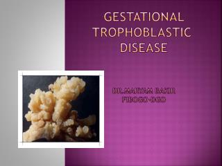
Hydatidiform mole Complete moles Hydropic degeneration of all villi Villous edema, trophoblastic hyperplasia, fetal-derived blood vessels disappear in stroma Partial moles Embryo or fetus exist Villous edema partially, trophoblastic proliferation lighterly, fetal-derived blood vessels presentin stroma
Partial moles Complete moles
Hydatidiform mole Related Factors • Complete moles • Area common in Latin America, Asia uncommon in North America and Europe • Race differences of the same race in different regions • Nutrition and Economylack of Vit A • Age < 20 or >35 years • Chromosome karyotype: Diploid • an empty egg fertilized with 1 haploid sperm(46,XX) 90% • an empty egg fertilized with 2 haploid sperm(46,XY) 10% • Genomic imprinting disorder NLRP mutation (paternal imprint gene deficiency)
Hydatidiform mole • Partial moles • high-risk factors are still unknown • "Haploid egg" fertilization usually two sperm fertilize a normal egg a triploid karyotype (69 chromosomes ), with the extra haploid set of chromosomes derived from father
Hydatidiform mole Clinical Manifestations • Complete moles • Abnormal vaginal bleeding during early pregnancy( 8-12week) most common symptom • Uterine enlargement exceeding normal pregnant uterus • Others Abdominal pain Pregnancy-induced hypertension Theca lutein ovarian cyst Hyperthyroidism (CHM) • Partial moles • Symptoms mild, might misdiagnosed as abortion
Hydatidiform mole hCG regression pattern after hydatidiform Time of hCG regress to normal —9 weeks, no more than 14 weeks Abnormal hCG regression pattern after hydatidiform signifies the presence of GTN • Complete mole • 15% local invasion and 4% distant metastasis • High –risk factors: ① HCG>100,000U/L ② Enlargement of Uterine ③ Theca lutein ovarian cyst >6cm • Partial mole • 4% local invasion and almost no distant metastasis • High –risk :unclear
Hydatidiform mole • Diagnosis • Abnormal bleeding after amenorrhea • Inappropriately enlarged uterus • Absence of fetal heart sounds not palpate fetus between 16-20th week • Vaginal discharge hydatidiform-like tissue Hydatidiform mole should be considered
Hydatidiform mole Diagnosis Ultrasound Complete moles produce a characteristic sonographic pattern, usually referred to as a “snowing” pattern HCG Elevated level exceed expected depending gestational age Continue to rise after 8-10 weeks of gestation HCG-related molecules Hyperglycosylated HCG free β-HCG subunit DNA karyotype Complete moles — usually diploid Partial moles — usually triploid
Hydatidiform mole • Treatment Suction curettage • Molar pregnancy should be terminated as soon as possible when diagnosis has been confirmed • Suction curettage is a first choice, must be done in operating room • Samples should be submitted to pathology
Hydatidiform mole Treatment • Theca lutein cysts of the ovary do not need treatment • Prophylactic chemotherapy: A controversial topic only be offered to patients with high-risk factor or with difficulty for follow-up • Hysterectomy Couldn’t prevent distant metastasis Only for old women without childbearing desire
Hydatidiform mole Follow-up • necessary for diagnosis of early GTN • Methods: • HCG • Symptom: Abnormal uterine bleeding • Pelvic examination • Ultrasound, chest X-ray and CT • Contraception: • Condom and oral contraceptives, not IUD • Duration for contraception — 1 year
General Consideration Antecedent gestation 60% hydatidiform mole 30% follow abortion 10% term pregnancy or ectopic pregnancy from mole —invasive mole or choriocarcinoma from Non-mole —choriocarcinoma
Gestational Trophoblastic Neoplasia Pathology • Invasive mole • Hydatidiform mole that invades the myometrium and may produce distant metastases • Microscopic finding are the same as in hydatidiform mole • Choriocarcinoma • Gloss:invades the myometrium , penetrate the serosa and may produce distant metastases • Microscopy:no villi, but instead sheets or foci of trophoblasts on a background of hemorrhage and necrosis
Invasive mole Choriocarcinoma Invasive mole Choriocarcinoma Invasive mole Choriocarcinoma
invades the myometrium Lung metastases Brain metastases cervical metastases
Gestational Trophoblastic Neoplasia Clinical Manifestation Nonmetastatic GTN • Antecedent gestational event is usually HM • Abnormal vaginal bleeding after mole • Others: • Enlarged uterus • Theca lutein cysts of the ovary • Abdominal pain • Fake pregnancy symptoms
Gestational Trophoblastic Neoplasia Metastatic GTN Usually chroriocarcinoma • Primary symptoms • Metastatic symptoms • lung metastases are frequently common • vaginal metastases are the second • liver and brain metastases usually death cause • other metastastic sites spleen, kidney, bladder, gastrointestinal system, and bone Simultateously occur or not
Gestational Trophoblastic Neoplasia Diagnosis • Symptoms and signs: ◆ Abnormal vaginal bleeding after post-evacuation, abortion, term pregnancy or ectopic pregnancy, ◆ Metastatic symptoms GTN should be considered
Gestational Trophoblastic Neoplasia • HCG assay Most important and main diagnostic evidence Diagnostic criteria for post- HM GTN hCG plateau for >4 values (±10%), over 3 weeks hCG increase of ≥10% over 2 weeks Diagnostic criteria for non post-HM GTN HCG elevated at 4w after abortion, term or ectopic pregnancy Re-rising HCG titer after reaching normal levels
Gestational Trophoblastic Neoplasia • Chest X-ray lung metastases • CT small lung metastases and brain metastases • MRI Liver and brain metastases • Ultrasound primary lesions of uterus and pevical metastases Imaging supports diagnosis, but not necessary
Gestational Trophoblastic Neoplasia • Histological diagnosis • villus shape can be found in primary or metastatical lesions • Presence of villus shape Invasive mole Absence of villus shape Choriocarcinoma Histology is not necessary for diagnosis of GTN
Anatomy staging of GTN (FIGO, 2000) Gestational Trophoblastic Neoplasia Stage III Stage I Stage II Stage IV
Prognostic scoring system for GTT (FIGO,2000) * Total score≤6 low risk, ≥7 high risk
Gestational Trophoblastic Neoplasia Treatment • Chemotherapy combining surgery, radiotherapy and other treatment • Stratified base on the prognostic scoring and stage Chemotherapy : • Single-agent chemotherapy is applied in low-risk gestational trophoblastic disease (MTX, Act-D, 5-Fu) • High-risk patients commonly use combined chemotherapy (EMA-CO)
Single agent chemotherapy DAY Therapy Interval 1-5 MTX 0.4mg/kg im qd 14d 1、3、5、7 MTX1mg/kg im 14d 2、4、6、8 FA 0.1mg/kg im or po 1-5 Act-D10-12ug/kg ivgtt qd 14d 1-8 5-Fu 28-30mg/kg ivgtt qd 12-14d
PSTT • A special type, more rarely in clinic • Most of them have a good prognosis • From intermediate trophoblast cells • Clinical manifestations • More common occur at reproductive period women • More common occur following term pregnancy • Abnormal bleeding after amenorrhea
PSTT • Diagnosis HCG negative or mildly elevated HPL mildly elevatedConfirmed by histology • Treatment Surgery is the preferred treatment Chemotherapy is adjuvant therapy