Download
1 / 58
580 likes | 607 Views
Skeletal system. An Introduction to the Skeletal System. The Skeletal System Includes: Bones of the skeleton Cartilages, ligaments, and connective tissues. Functions of the Skeletal System. Five Primary Functions of the Skeletal System Support

E N D
An Introduction to the SkeletalSystem • The SkeletalSystem • Includes: • Bones of theskeleton • Cartilages, ligaments, and connectivetissues
Functions of the SkeletalSystem • Five Primary Functions of the SkeletalSystem • Support • Storage of Minerals (calcium) and Lipids (yellow marrow) • Blood Cell Production (redmarrow) • Protection • Leverage (force ofmotion)
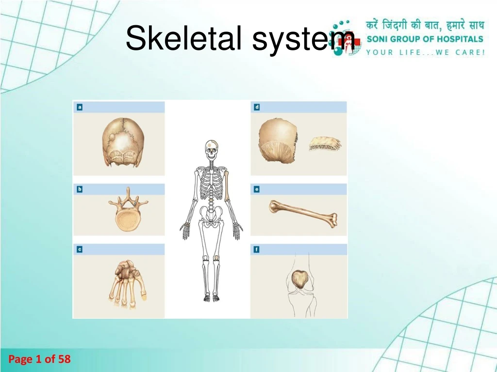
TheAxialSkeleton • Forms the longitudinal axis of thebody • Has 80bones • Theskull • 8 cranialbones • 14 facialbones • Bones associated with the skull • 6 auditoryossicles • The hyoidbone
The AxialSkeleton • The vertebralcolumn • 24 vertebrae (singular =vertebra) • Thesacrum • Thecoccyx • The thoraciccage • 24ribs • Thesternum
The AxialSkeleton Skull Cervical vertebrae Sternum Thoracic vertebrae Ribs Costal cartilages Lumbar vertebrae Sacrum Coccyx Anterior (left) and posterior (right) views of the axial skeleton. The individual bones associated with the skull are notvisible.
An Introduction to the AppendicularSkeleton • TheAppendicularSkeleton • 126bones • Allows us to move and manipulateobjects • Includes all bones besides axialskeleton • Thelimbs • The supportivegirdles
AppendicularSkeleton SKELETALSYSTEM 206 APPENDICULARSKELETON 126 AXIALSKELETON 80 (see Figure7–1) 2 Clavicle Pectoral girdle 4 Scapula2 Humerus2 Upper limbs 60 Radius2 Ulna 2 Carpal bones 16 Metacarpal 10 bones Phalanges28 Pelvic girdle Hipbone2 2
ThePelvicGirdle • Made up of two hip bones (coxalbones) • Strong to bear body weight, stress ofmovement • Part of thepelvis • Coxalbones • Made up of three fusedbones • Ilium (articulates withsacrum) • Ischium • Pubis
The Appendicular Skeleton (Part 2 of2) Femur 2 Lower limbs 60 Patella 2 Tibia 2 2 Fibula Tarsal bones14 Metatarsal bones 10 Phalanges 28
Classification ofBones • Bones • Are classifiedby: • Shape • Internal tissueorganization • Bone markings (surface features;marks)
Classification ofBones • Six BoneShapes • Suturalbones • Irregularbones • Shortbones • Flatbones • Longbones • Sesamoidbones
Classification of Bones byShape FlatBones SuturalBones Sutures Externaltable Parietalbone Sutural bone Internal table Diploë (spongy bone) LongBones IrregularBones Vertebra Humerus ShortBones SesamoidBones Patella Carpal bones
An Introduction to the AxialSkeleton • Structures ofBones • Articulations • Contacts with otherbones • Landmarks (bone markings;marks) • Areas of muscle and ligamentattachment • Foramina • Openings for nerves and bloodvessels
Classification ofBones • Bone Markings • Depressions orgrooves • Along bonesurface • Elevations orprojections • Where tendons and ligamentsattach • At articulations with otherbones • Tunnels • Where blood and nerves enterbone
An Introduction to Bone Markings Trochanter Head Neck Sinus Head Sulcus Tubercle Crest Neck Fossa Foramen Fissure Spine Process Tuberosity Ramus Line Facet Tubercle Foramen Fossa Ramus Trochlea Pelvis Skull Condyle Humerus Condyle Femur
Classification ofBones • Structure of a Long Bone • Diaphysis • Theshaft • A heavy wall of compact bone, or densebone • A central space called medullary (marrow)cavity • Epiphysis • Wide part at eachend • Articulation with otherbones • Mostly spongy (cancellous)bone • Covered with compact bone(cortex) • Metaphysis • Where diaphysis and epiphysismeet
Epiphysis Spongy bone Metaphysis BoneStructure Compact bone Diaphysis (shaft) Medullary cavity Metaphysis Epiphysis The structure of a representative long bone (the femur) in longitudinalsection
Bone (Osseous)Tissue • Bone (Osseous)Tissue • Dense, supportive connectivetissue • Contains specializedcells • Produces solid matrix of calcium saltdeposits • Around collagenfibers
Bone (Osseous)Tissue • Characteristics of Bone Tissue • Dense matrix,containing: • Deposits of calciumsalts • Osteocytes (bone cells) within lacunae organized around bloodvessels • Canaliculi • Form pathways for bloodvessels • Exchange nutrients andwastes
Bone (Osseous)Tissue • Characteristics of Bone Tissue • Periosteum • Covers outer surfaces ofbones • Consists of outer fibrous and inner cellularlayers
Bone (Osseous)Tissue • BoneMatrix • MatrixProteins • One third of bone matrix is protein fibers(collagen)
Compact Bone andSpongyBone • The Structure of CompactBone • Osteon is the basicunit • Osteocytes are arranged in concentriclamellae • Around a central canal containing bloodvessels • Perforatingcanals • Perpendicular to the centralcanal • Carry blood vessels into bone andmarrow
The Blood Supply to a MatureBone Articularcartilage Epiphyseal artery andvein Branches of nutrientartery andvein Metaphyseal artery and vein Periosteum Periosteum Compact bone Medullary cavity Periosteal arteriesand veins Connections tosuperficial osteons Nutrientartery andvein Nutrientforamen Metaphysis Metaphyseal artery andvein Epiphyseal line
The Structure of CompactBone Circumferential lamellae Venule Capillary Periosteum Osteons Perforating fibers Interstitial lamellae Concentric lamellae Trabeculaeof spongy bone (seeFig.6–6) Vein Perforating canal Artery Arteriole Central canal The organization of osteonsand lamellae in compactbone
Bone (Osseous)Tissue • BoneMatrix • Minerals • Two thirds of bone matrix is calcium phosphate, Ca3(PO4)2 • Reacts with calcium hydroxide,Ca(OH)2 • To form crystals of hydroxyapatite, Ca10(PO4)6(OH)2 • Which incorporates other calcium salts andions
Bone Landmarks for Physicalexaminaiotn • Most prominent spinous process isC7/T1. • Inferior points of scapulae are at interspace between T7 &T8. • Superior margin of each iliac crest crossesL4. • Two symmetric dimples overlying posterior superior iliac spine are at level ofS2. • Coccyx.
The AppendicularSkeleton SKELETALSYSTEM 206 APPENDICULARSKELETON 126 AXIALSKELETON80 2 Clavicle Pectoral girdle 4 Scapula2 Humerus2 Upper limbs 60 Radius2 Ulna 2 Carpal bones 16 Metacarpal 10 bones Phalanges28 Pelvic girdle Hipbone2 2
Physical ExaminationLandmarks • You should be able to palpate these: • Elbow (lateral and medial epicondyles of humerus, olecranon process of ulna) • Wrist (styloid process of radius – thumb side, & of ulna) • Shoulder (acromian process ofscapula) • Hip (iliac crest, greater trochanter offemur) • Knee(patella) • Shin (anterior crest of tibia) • Ankle (lateral malleolus of fibula & medial malleolus oftibia) • Heel(calcaneus).
Articulations Prepared by DidyButton
An Introduction toArticulations • Articulations • Body movement occurs at joints (articulations) where two bonesconnect • JointStructure • Determines direction and distance of movement (range of motion orROM) • Joint strength decreases as mobilityincreases
Classification ofJoints • FunctionalClassifications • Synarthrosis (immovablejoint) • Amphiarthrosis (slightly movablejoint) • Diarthrosis (freely movablejoint)
Classification ofJoints • Structural Classifications • Fibrous joints. Articulating bones bound tightly together by fibrous connective tissue (sutures, syndesmosis between tibia & fibula,gomphoses). • b) Cartilaginous joints. Articulating bones held tightly together by cartilage (synchondroses between rib & sternum, pubicsymphysis). • c) Synovial joints. Articulating bones have a fluid-filled space between them (gliding, hinge, pivot, ellipsoidal, saddle, ball &socket).
SynovialJoints • SynovialFluid • (e.g. knee, hip, elbow, shoulder, fingers, toes,jaw) • Contains slippery proteoglycans secreted byfibroblasts • Functions of synovialfluid • Lubrication • Nutrientdistribution • Shockabsorption
The Structure of a SynovialJoint Medullarycavity Spongybone Periosteum Fibrous jointcapsule Synovial membrane Articularcartilages Joint cavity (containing synovialfluid) Compactbone Synovial joint, sagittalsection
SynovialJoints • AccessoryStructures • Cartilages • Fatpads • Ligaments • Tendons • Bursae
SynovialJoints • Factors That Stabilize SynovialJoints • Prevent injury by limiting range ofmotion • Collagen fibers (joint capsule,ligaments) • Articulating surfaces andmenisci • Other bones, muscles, or fatpads • Tendons of articulatingbones
The Structure of a SynovialJoint Quadricepstendon Bursa Jointcapsule Patella Articularcartilage Femur Synovial membrane Meniscus Fatpad Patellarligament Intracapsular ligament Jointcavity Tibia Meniscus Knee joint, sagittalsection
Inflammation of the synovialjoint • Osteoarthritis bony growths (osteophytes) at joints • Rheumatoid arthritis is an autoimmune disease characterised by chronic inflammation of the synovial membrane leading to deformity, loss of function, pain, swelling, ultimately to joint destruction. • Psoriatic arthritis associated with the skin condition psoriasis.
Figure 9-3a AngularMovements Extension Flexion Hyperextension Flexion Hyper- extension Flexion Extension Extension Flexion Hyperextension Extension Flexion/extension
AngularMovements Abduction Abduction Adduction Adduction Abduction Adduction Abduction Adduction Abduction/adduction
AngularMovements Abduction Adduction Adduction/abduction
AngularMovements Circumduction
RotationalMovements Headrotation Right rotation Left rotation Lateral (external) rotation Medial (internal) rotation
Rotational Movements Pronation Supination Supination Pronation
Figure 9-5 SynovialJoints Inversion Eversion
Figure 9-5 SynovialJoints Dorsiflexion (ankleflexion) Plantar flexion (ankleextension)
The KneeJoint • Seven Major SupportingLigaments • Patellar ligament(anterior) • & 3. Two popliteal ligaments(posterior) • 4. & 5. Anterior and posterior cruciate ligaments (inside jointcapsule)
Tibia collateral ligament(medial) Fibular collateral ligament(lateral)
Figure 9-12a The Right Knee Joint Quadriceps tendon Patella Joint capsule Patellar retinaculae Tibial collateral ligament Fibular collateral ligament Patellar ligament Tibia Anterior view, superficial layer