Download

1 / 11
160 likes | 1.14k Views
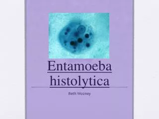
Entamoeba histolytica Pathogenesis and pathology . Factors that may influence the pathogenesis: 1- strain variation. 2- role of bacteria . 3- infective dose . 4- nutritional status . 5- associated disease. 6- pregnancy. 7- drugs. 8- intestinal mucus. 9- dietary iron.

E N D
EntamoebahistolyticaPathogenesis and pathology Factors that may influence the pathogenesis: 1-strain variation. 2-role of bacteria. 3-infective dose. 4-nutritional status. 5-associated disease. 6-pregnancy. 7-drugs. 8-intestinal mucus. 9-dietary iron.
Characters of intestinal amoebic ulcer -Size:pinhead to one inch -Shape:flask-shaped, water bottle -Margins:ragged -Distribution: along the whole length of L.I ileo-caecalor sigmoido-rectal region -Extension of the ulcer: superficial ulcers deep ulcers -Healing: without any scar with scar
Clinical features -IP: 1-4 weeks. 1- primary or intestinal amoebiasis. 2- secondary or extra-intestinal amoebiasis.
Intestinal amoebiasis • -amoebic dysentery • -non-dyseteric amoebic colitis. • -amoebic appendicitis. • -amoeboma. • -complications.
-Complications of intestinal amoebiasis a-Local complications: • 1-intestinal perforation. • 2-peritonitis. • 3-haemorrhage. • 4-stricture and obstruction. • 5-prolapse of rectum. • 6-intussusception.
Extraintesinalamoebiasisb-Systemic complications: • -amoebic hepatitis. • -amoebic liver abscess. • -pulmonary amoebisis. • -cerebral amoebisis. • -cuteneousamoebiasis. • -splenic abscess
Amoebic liver abscess Gross pathology of amoebic liver abscess: -Size: 10-15 cm in diameter -location: right lobe -Size of liver: enlarged -Cut section: liquefaction of the central necrotic -Color of pus: anchovy sauce like pus -Wall of the abscess: ragged
Microscopic pathology of amoebic liver abscess 1-central zone: cytolysed material with no amoebae. 2-intermediate zone: degenerated liver cells, red blood cells, white blood cells, connective tissue and occasionally trophozoites 3-peripheral zone: congested capillaries varying degree of necrosis of liver cells well demonestratedtrophozoites