Download
1 / 83
1.13k likes | 3.56k Views
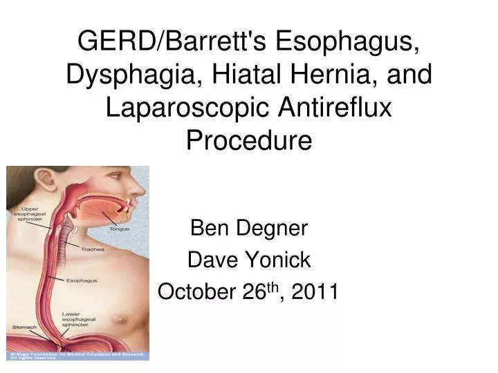
GERD/Barrett's Esophagus, Dysphagia, Hiatal Hernia, and Laparoscopic Antireflux Procedure . Ben Degner Dave Yonick October 26 th , 2011. Anatomy. Mucosa, submucosa, muscularis propria, and adventitia Lack serosa vs. other GI tract Mucosa innermost (4 layers) contains squamous epithelium.

E N D
GERD/Barrett's Esophagus, Dysphagia, Hiatal Hernia, and Laparoscopic Antireflux Procedure Ben Degner Dave Yonick October 26th, 2011
Anatomy • Mucosa, submucosa, muscularis propria, and adventitia • Lack serosa vs. other GI tract • Mucosa innermost (4 layers) • contains squamous epithelium
Muscularis Propria • Continuation of inferior constrictor of the pharynx • Two muscle bundles • inner circular • outer longitudinal • Striated upper 1/3 -vagus and its recurrent laryngeal branches • Smooth lower 2/3 -visceral nerve plexus derived from neural crest cells • Left vagus anterior-liver/biliary tree • Right vagus posterior-celiac plexus
Esophageal Arteries Upper-Inferior Thyroid Artery Lower-Left Gastric and Inferior Phrenic arteries, bronchial arteries and 4-6 aortic branches
Esophageal Lyphatics Lymphatics: upper 2/3 cephalad, lower 1/3 caudad
Anatomic Areas of Narrowing • Cricopharyngeal muscle • Left mainstem bronchus and aortic arch • Diaphragm
UES • 15 cm from incisors • Cricophayrngeus muscle, recurrent laryngeal nerve • Site of perforation is cricopharyngeus muscle (with EGD), aspiration if UES fail
LES • 40 cm from incisors • No anatomic landmarks • rise in pressure when transducer is pulled from the stomach • Increased Pressure: Alpha-adrenergics, BBs, gastrin, motilin, antacids, cholinergics, metoclopramide • Decreased Pressure: Alpha blockers, Beta andrenergics, CCK, estrogen, glucagon, progesterone, somatostatin, secretin, barbiturates, CCBs, caffeine, diazepam, dopamine, meperidine, ethanol, coffee, fat
LES • High pressure except: 1. passage of food into the stomach 2. when fundus is distended with gas, LES eliminated to allow venting of the gas • Loss of the normal high-pressure zone leads to GERD (transient vs permanent)
Gastroesophageal Reflux Disease • 1/3 Western population experience symptoms at least once a month • 4-7% daily • Most patients with mild symptoms carry out self-medication • The prevalence and severity of GERD is increasing
Typical GERD Symptoms • Heartburn • substernal burning or chest pain • worse with spicy foods, tomato sauce, citrus juices, chocolate, coffee, and alcohol • 1 to 2 hours after eating, often at night, relieved by antacids and OTC H2 blockers • Regurgitation • sensation that fluid or food is returning into the esophagus • worse at night or when lying down after a meal • Dysphagia • up to 40% of pts with GERD have sensation of food hanging up in the lower esophagus--esophageal dysphagia • typically limited to only solid food, with normal passage of liquids, suggesting mechanical disorder • develops slowly enough that the patient may adjust eating habits unknowingly
Atypical GERD Symptoms • Cough, asthma, hoarseness, and noncardiac chest pain • primary complaint in 20-25% • more difficult to prove a cause-and-effect relationship • trial of high-dose PPIs is helpful • make sure patient doesn’t have another cause for pain
Pathophysiology of GERD • Fundic distention because of overeating • LES is taken up by the expanding fundus, exposing the squamous epithelium/LES to gastric juice • Worsened by delayed gastric emptying with high-fat diet and hiatal hernia • Compensated with increased swallowing • Saliva bathe the injured mucosa and alleviate the discomfort = aerophagia, bloating, repetitive belching • More distension leads to further exposure and repetitive injury to the terminal squamous epithelium leading to inflammation • continued epigastic pain and possibly epithelial columnarization • Extension of the inflammatory process into the muscularis propria • leading to a permanently defective sphincter
Diagnosis of GERD • Based on symptoms alone? • Correct in only 2/3 of patients • these symptoms are not specific for GE reflux • achalasia, diffuse spasm, esophageal carcinoma, pyloric stenosis, cholelithiasis, gastritis, gastric or duodenal ulcer, and coronary artery disease • need objective diagnosis before the decision is made for surgical treatment
Diagnosis of GERD • First episode • Initial therapy with H2 blockers or PPI for 12 weeks • Failure of H2 blockers or PPI to control the symptoms suggests that either the diagnosis is incorrect or the patient has severe disease • EGD • Opportunity for assessing the severity of mucosal damage • 24-hour pH and bilirubin monitoring • Measurement degree and pattern of esophageal exposure to gastric and duodenal juice • Manometry • Assess the status and function of the LES and esophageal body • These studies identify features that predict a poor response to medical therapy, frequent relapses, and the development of complications
Complications of GERD • Mucosal complications-esophagitis and stricture • Extraesophageal or Respiratory complications, such as laryngitis, recurrent pneumonia, and progressive pulmonary fibrosis • Reflux (aspiration) vs reflex (vagal bronchoconstriction) • Metaplastic and Neoplastic complications, Barrett's and esophageal adenocarcinoma • Prevalence/severity of complications related to the degree of loss of the GE barrier and content of refluxed gastric juice, not symptoms
Barrett’s Esophagus • Squamous epithelium metaplasia columnar epithelium • 7-10% of patients with GERD • Normally, the SCJ should coincide with the GEJ (linear gastric mucosal folds) • Presence of any columnar mucosa extending at least 3 cm into the esophagus (goblet cells)=Barrett’s • predisposed to malignant degeneration • Increased risk of adenocarcinoma x50
Classification and Management of Barrett’s Esophagus with Dysplasia • Indefinite for Dysplasia: Aggressive antireflux therapy (60 mg PPI per day) and repeated biopsy in 3 months • Low Grade: Aggressive antireflux therapy vs. surgical treatment • High Grade-Esophagectomy and PPI
Dysphagia Difficulty in transferring a food from the mouth to the stomach Regurgitation, chest pain, heartburn, and coughing or choking spells Oropharyngeal functional disturbance in the swallowing mechanism Esophageal mechanical obstruction or esophageal motility disorder
Dysphagia Evaluation of a patient with dysphagia must be performed in a systematic manner Barium swallow Additional diagnostic tests EGD, manometry, 24-hour pH study, and possibley bronchoscopy and endoscopic ultrasonography (EUS). Diagnostic imaging by CT and PET in assessing patients with esophageal cancer
Oropharyngeal Dysphagia inability to chew food, drooling, coughing during a meal, and nasal regurgitation of solids or liquids dysphagia within 1 second of swallowing The common causes can be grouped into three broad categories: 1) generalized systemic conditions: CVA, Myasthenia gravis 2) intrinsic functional disturbances: Zenker's diverticulum 3) fixed mechanical obstruction: Neoplasm, webs, previous surgical treatment, previous radiation therapy
Esophageal Dysphagia Dysphagia with solids? =Mechanical Obstruction Intermittent? Esophageal Ring or Esophagitis Progressive with GERD? Peptic Stricture Progressive with weight loss and anorexia? Esophageal Cancer Dysphagia for both liquids and solids? =Motility Disorder Intermittent? Spasm (DES) Progressive with GERD? Scleroderma Progressive? Achalasia
Schatzki's Ring symmetrical narrowing at SCJ, small hiatal hernia correlation with GERD barium swallow and esophagoscopy to confirm Asymptomatic? no specific treatment is needed Definitive treatment? dilatation of the ring with medical therapy for GERD. If refractory, dilatation plus antireflux surgery
Peptic Stricture H/o GERD worsening dysphagia for years without weight loss End stage of ulcerative esophagitis, healing ulcer causes annular fibrosis Dx: barium swallow followed by upper GI endoscopy greater length and more tapered than Schatzki’s
Esophageal Webs localized narrowing of the esophagus caused by intraluminal extension of the mucosa and part of the submucosa congenital or acquired (mc), usually secondary to conditions such as iron deficiency anemia/Plummer-Vinson syndrome and ulcerative colitis. Tx: endoscopic dilatation
Achalasia Dysphagia for liquids and solids and possibly weight loss. Barium swallow shows absent peristalsis and a dilated esophagus, possibly tapered narrowing in distal esophagus=bird's beak Achalasia risk factor for squamous cell cancer Tx: Pneumatic dilatation or surgery
Diffuse esophageal spasm unknown etiology Nonprogressive dysphagia with solids and liquids and nonexertional chest pain that responds to nitroglycerin corkscrew on barium The diagnosis by manometry periodic occurrence of simultaneous high-amplitude contractions with intervening periods of normal peristalsis. Tx: r/o CAD, then medical management of reassurance, nitrates, and CCBs Botulinum toxin injection, surgery does not have an established role
Nutcracker Esophagus unknown etiology women>men Manometry: peristaltic waves with significantly elevated amplitude (> 180 mm Hg). Treatment is primarily medical
Hypertensive LES unknown etiology can occur alone or in association with achalasia, nutcracker esophagus, or DES Manometry-LES pressure over 45 mm Hg Tx: primarily medical, but balloon dilatation is done
Esophageal Diverticula < 5% of all cases of dysphagia. False diverticula (pulsion) include only the mucosal layer underlying motor dysfunction True diverticula (traction) include all layers of the esophageal wall inflammatory process Esophageal diverticula may also be classified into three categories on the basis of the anatomic level at which they occur
Pharyngoesophageal/Zenker’s Diverticula MC from muscle incoordination that leads to herniation of the mucosa in prox esophagus Dysphagia mc symptom, halitosis, regurgitation, throat discomfort, palpable neck mass, recurrent aspiration pneumonia The best initial diagnostic tool is a barium swallow perforation in EGD
Midesophageal diverticula True traction diverticula Caused by periesophageal inflammation in granulomatous inflammation of the subcarinal lymph nodes from TB or fungal infection Frequently asymptomatic and are often found incidentally Dysphagia does occur but is a rare symptom
Epiphrenic diverticula Acquired pulsion diverticula of distal esophagus Associated with other esophageal motor disorders (achalasia, DES, and hypertensive LES) but can occur alone Absent or mild symptoms? Conservative management is appropriate Significant dysphagia? Surgical management
Chemical Ingestion Alkali household cleaning agents Most occur accidentally in children, but suicide in adults Magnitude and site of the injury? Related to the length of the contact time Injury at any level, MC is distal esophagaus lead to submucosal scar formationstricture and dysphagia Endoscopic exam is first step A barium swallow should be done in the first month after injury to detect any stricture and then serial swallows
Hiatal Hernia I-Sliding, dilation of hiatus, most commonly associated with GERD -most with reflux have sliding, most with sliding don’t have reflux II-Paraesophageal, defect in diaphragm alongside esophagus with normal GE junction --chest pain, dysphagia, early satiety III-Combined I and II IV-entire stomach in chest plus another organ (colon, spleen)
Laparoscopic Antireflux Procedure • Most commonly performed procedure is a fundoplication • Nissen Fundoplication: 360 degree fundoplication • Laparoscopic approach reduces postoperative pain and shortens length of hospital stay • Rapid increase in surgical treatment of GERD
Preoperatively • h/o recurrent heartburn enough to clinically establish GERD and initiate empiric medical therapy • Those patients with recurrent or refractory symptoms require further evaluation prior to surgery • Endoscopy • Evaluate for Barrett’s Esophagus • Manometry • Evaluate for other causes of esophageal dysmotility • 24 hour pH • May be helpful, especially in those patients with atypical GERD symptoms or other GI comorbidities
Indication • Failure of maximal medical therapy • Short 2 month trial • Failure of lifestyle modification • Weight loss • Alteration in diet (avoid chocolate, peppermint, fat, onions, garlic, alcohol, caffeine, nicotine) • Avoid food 2-3 hours prior to sleep • Elevation of head of bed 6 – 10 inches • Limit potentially precipitating activities (bending over or strenuous exercise) • Complications of GERD • Esophagitis, stricture, recurrent aspiration or pneumonia, Barrett’s esophagus • Associated with paraesophageal hernia • Intolerance to medical therapy or patient desiring discontinueation of medical therapy
Contraindication • Absolute • Inability to tolerate general anesthetic or laparoscopy • Uncorrectable coagulopathy • Relative • Previous upper abdominal surgery • Morbid obesity • Severe esophagitis with or without stricture • Small-body habitus • Short esophagus • Particularly for fundoplications • Paraesophageal hiatal hernia
Basic tenets of antireflux surgery • Restoration of an effective LES • Creation of a gastroesophageal valve • Mechanical effects of a fundoplication • Fundus exhibits a physiologic phenomenon of receptive relaxation • Decreased tone of the gastric fundic smooth muscle in association with swallowing-induced relaxation of the LES
Belsey Mark IV Repair • Transthoracic repair to control GERD • Typically performed through a left thoracotomy • May be performed thoracoscopically • McKernan and Champion modified Belsey • Distal esophagus and proximal stomach are mobilized and ddelivered through the esophageal hiatus • Anterior 270 degree plication of the fundus is performed onto the esophagus • The fundoplication is buttressed by the diaphragmatic crura
Nissen Fundoplication • 360 degree wrap around esophagus • Circumferentially dissecting the distal esophagus • Mobilizing the gastric fundus • Plicating the fundus around the lower esophagus • Creating a high-pressure zone • Increases the resting tone of the sphincter mechanism • Improves its response to elevated intragastric pressure • Fundoplication varied from 3 – 6 cm and included the esophageal wall in the fundoplication • No division of the short gastric vessels
Rossetti-Hell modification • Included wrapping the anterior portion of the fundus around the esophagus • No division of the short gastric vessels • Minimal moblization of the upper stomach • Created a 3 – 6 cm 360 degree fundoplication
Short, floppy Nissen • Most common modification used today • The short gastric vessels are divided • Full fundic mobilization and the lateral border of the fundus is wrapped around the esophagus • Sutured to the medial edge of the medial fundus • Short fundoplication, < 2.5 cm • Fashioned over a 50 to 60F bougie