Download

1 / 42
440 likes | 639 Views
Histopathologie de la Borréliose de Lyme. Dr Moguelet Service d’Anatomie pathologique CHU TENON. Histopathologie . Réponse cellulaire inflammatoire Lymphocytes B et T, plasmocytes , macrophages et mastocytes Biopsie cutanées / synoviales / musculaires. Histopathologie.

E N D
Histopathologie de la Borréliose de Lyme Dr Moguelet Service d’Anatomie pathologique CHU TENON
Histopathologie • Réponse cellulaire inflammatoire • Lymphocytes B et T, plasmocytes, macrophages et mastocytes • Biopsie cutanées / synoviales / musculaires
Histopathologie • Les lésions cutanées • Les lésions articulaires • Les lésions musculaires • Les lésions cardiaques • Les lésions neurologiques
Histopathologie • Image évocatrice • Si plasmocytes présents • Non spécifique • Diagnostic d’élimination • Nombreux diagnostics différentiels
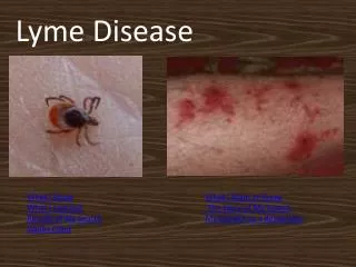
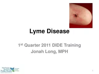
Les lésions cutanées • Site de piqûre de tique • Erythème migrant • Lymphocytome cutané bénin • Acrodermatite chronique atrophiante
Diagnostics différentiels • Les lésions cutanées • Site de piqûre de tique • Piqûre d’insecte • Erythème migrant • Lymphocytome cutané bénin • Acrodermatite chronique atrophiante
Les lésions cutanées • Site de piqûre de tique • Erythème migrant • Lymphocytome cutané bénin • Acrodermatite chronique atrophiante
Diagnostics différentiels • Les lésions cutanées • Site de piqûre de tique • Erythème migrant • Érythèmes annulaires • Lymphocytome cutané bénin • Acrodermatite chronique atrophiante
Diagnostics différentiels • Les lésions cutanées • Site de piqûre de tique • Erythème migrant • Érythèmes annulaires • Lymphocytome cutané bénin • Acrodermatite chronique atrophiante Erythema annulare centrifugumErythema gyratum repensErythema chronicum migransErythema marginatumAnnular erythema of infancy
Les lésions cutanées • Site de piqûre de tique • Erythème migrant • Lymphocytome cutané bénin • Acrodermatite chronique atrophiante
Diagnostics différentiels • Les lésions cutanées • Site de piqûre de tique • Erythème migrant • Lymphocytome cutané bénin • lymphome B cutané • Pseudolymphome B • Acrodermatite chronique atrophiante
Lymphocytome cutané bénin Lymphome B centrofolliculaire
Les lésions cutanées • Site de piqûre de tique • Erythème migrant • Lymphocytome cutané bénin • Acrodermatite chronique atrophiante
Diagnostics différentiels • Les lésions cutanées • Site de piqûre de tique • Erythème migrant • Lymphocytome cutané bénin • Acrodermatite chronique atrophiante • morphée • lichen scléroatrophique • DIG
Les lésions articulaires • synovite hypertrophique non spécifique
Diagnostics différentiels • synovite hypertrophique non spécifique • Polyarthrite rhumatoïde • Syndrome de Reiter • Synovite post-traumatique
Les lésions cardio-vasculaires • Cardiaque • 3 feuillets • infiltrat lymphoplasmocytaire • en bande sous endocardique • myocytes dégénératifs ou nécrotiques • microangiopathie oblitérante • Peau / synoviale / cœur / nerfs Duray PM. 1999
Les lésions musculaires • infiltrat lymphoplasmocytaire focal • interstitiel et périvasculaire • +/- fibres musculaires dégénératives • +/- fasciite
Les lésions du système nerveux • système nerveux central / méninges • Lymphocytes+++ • système nerveux périphérique • infiltrat lymphoplasmocytaire • Radiculaire : ganglionnaire dorsale • Perte de cellules ganglionnaires • Nerfs périphériques • Perte axonale multifocale
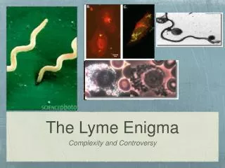
Méthode de visualisation in situ des Borrelia • Les colorations argentiques • Warthin Starry / Dieterle / Steiner / Bosma-Steiner • Réalisation difficile • Interprétation difficile, risques de faux positifs • Pas de différenciation des genres
N. Marti Ras, D. Postic, P. Ave, M. Huerre, G. Baranton. Res. Mic. 2000 (B. turicatae)
Méthode de visualisation in situ des Borrelia • Immunohistochimie • Ac mono-polyclonaux N. Marti Ras, D. Postic, P. Ave, M. Huerre, G. Baranton. Res. Mic. 2000 (B. turicatae)
Visualisation in situ • ACA : 10 à 120 coupes sont nécessaires (de Koning et al)
Conclusion : Intérêt histopathologie • Images évocatrices, non spécifiques • Mise en évidence de Borrelia difficilement réalisable en routine