Download
1 / 14
140 likes | 190 Views
A 24-year-old female hairdresser presented with a 5-day history of jaundice, right upper quadrant discomfort, and general malaise. Her stool and urine were of normal colour.

E N D
A 24-year-old female hairdresser presented with a 5-day history of jaundice, right upper quadrant discomfort, and general malaise. • Her stool and urine were of normal colour.
She had no past medical history and used no prescription drugs, OTC medications or NSAIDs, no nutritional supplements or herbal remedies, and no illicit drugs. • She had not recently had a course of antibiotics. There was no history of previous blood transfusion, contact with hepatitis, or previous jaundice, and she had no tattoos. There was no significant family history.
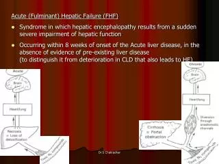
FULMINANT HEPATICFAILURE (FHF) • This is defined as severe hepatic failure in which encephalopathy develops in under 2 weeks in a patient with a previously normal liver. • Cases that evolve at a slower pace (2–12 weeks) are called subacute or subfulminant hepatic failure. • FHF is a rare but often life-threatening syndrome that is due to acute hepatitis from many causes
A 22-year-old male was admitted to hospital after being found by his landlord with confusion. • He was apyrexial, with a blood glucose of 5.4mmol/L, pulse rate 60 beats/min and regular, and blood pressure 114/58mmHg. • Heart sounds were normal and his chest was clear. He was noted to be jaundiced. • Asterixis was present. There were no focal neurological signs.
There were no spider naevi, muscle wasting, or gynaecomastia. His only medical history was pulmonary tuberculosis 6 months earlier, diagnosed by pleural biopsy and pleural fluid analysis. • He had last been followed up 2 months previously and was well at that time. He had completed 2 months of rifampicin, isoniazid, pyrazinamide and ethambutol, followed by rifampicin and isoniazid alone.
Investigations showed: • Hb 15.2g/dL, WBCs 9.1 x 10⁹/L, • Platelets 173 x 109/L • Na 135mmol/L, K 3.7mmol/L, urea 2.8mmol/L, creatinine 0.78mg/dl • Bilirubin 18.7mg/dl, ALT 1307 IU/L, ALP 436 IU/L, albumin 3.2g/dl • Prothrombin time 53.8 sec.
Clinical Features • Examination shows a jaundiced patient with a small liver and signs of hepatic encephalopathy. • The mental state varies from slight drowsiness, confusion and disorientation (grades I and II) to unresponsive coma (grade IV) with convulsions.
Fetor hepaticus is common, but ascites and splenomegaly are rare. • Fever, vomiting, hypotension and hypoglycaemia occur. • Cerebral oedema develops in 80% of patients with FHF but is far less common with subacute failure and its consequences of intracranial hypertension and brain herniation are the most common causes of death.
What blood tests would you request? A ‘liver screen’ should be performed to look for the cause of ALF including: ● Paracetamol concentration (although N-acetyl cysteine should be started before the result is known, to cover the possibility of toxicity). ● Hepatitis A IgM ● Hepatitis B core IgM ● Autoimmune markers: antinuclear antigen (ANA), anti-smooth muscle antibody (ASmAb), liver kidney microsomal antibody type 1 (LKM1), and immunoglobulins ● Copper and ceruloplasminto screen for Wilson’s disease.
What radiological tests would you request? • An ultrasound of the abdomen should be performed, since this will help distinguish between acute and chronic liver disease (splenomegaly is more common in chronic liver disease). • Ultrasound will not help to differentiate between different causes of ALF.
What is the management? • ● Intravenous rehydration: most patients require liberal volume expansion. • ● The blood glucose should be measured hourly and replaced: with intravenous glucose (10–50%) as necessary, since there is a high risk of hypoglycaemia
Arterial blood gas to monitor acidosis. • ● N-acetyl cysteine should be given to cover the possibility of paracetamol overdose. • ● Antibiotics: prophylaxis with an intravenous cephalosporin is recommended. Patients with ALF have a high risk of infection and sepsis from bacterial and/or fungal infection.
Vitamin K is not indicated: bleeding is unusual in ALF. • Fresh frozen plasma is not advocated, because the risks (fluid overload, noramalization of the prothrombin time artificially) outweigh the benefits. • ● Lactulose is of no proven benefit in ALF. • ● Intensive care management: if the patient is not protecting their airway • ● Liver transplant:


![]; Njhj;jpuk ; Jjpghj;jpuh ck;ik , d;Wk ; vd;Wk ; Jjpj;jpLNtd ;](https://cdn3.slideserve.com/6747820/slide1-dt.jpg)