Download
1 / 27
290 likes | 562 Views
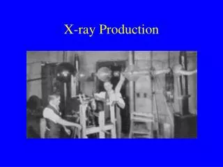
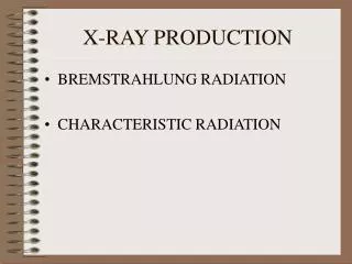
PREPARED BY Dr fahad albadr radiology chairman. X-RAY PRODUCTION AND EXPOSURE FACTORS. X-RAY PRODUCTION AND EXPOSURE FACTORES. IN 1895. Wilhelm Röntgen. What is X-Ray?.

E N D
PREPARED BY Dr fahadalbadr radiology chairman X-RAY PRODUCTION AND EXPOSURE FACTORS HABIS
X-RAY PRODUCTION ANDEXPOSURE FACTORES IN 1895 Wilhelm Röntgen H kkuh
What is X-Ray? • Is a high energy electromagnetic radiation produced when highly energetic electrons interact with matter • Travel at speed of light • Travels in a straight line Major usage of X-Ray • Medical Imaging • Diagnostic X-Ray Machine • Industrial Imaging • Airport Baggage Screening H kkuh
Electromagnetic Spectrum Visible Ionizing Radiation Nonionizing Radiation Infrared Ultraviolet Radar Near Far X Rays FM TV Gamma Rays Short wave Power Cosmic Rays Broadcast Transmission -14 -12 -10 -8 -6 -4 -2 2 4 6 8 10 10 10 10 10 10 10 10 10 10 10 1 Wavelength in Meters 10 8 6 4 2 -2 -4 -6 -8 -10 -12 -14 10 10 10 10 10 10 10 10 10 10 10 10 1 High Low Energy - Electron Volts HABIS
Nonionizing Radiation • Sources • Ultraviolet light • Visible lightMicrowaves • Infrared radiation • Radio & TV
Energy Wood Concrete Alpha Low Beta Medium X-RAY & Gamma High Ionizing Radiation
The X-ray tube parts: • Cathode (-) • Filament made of tungsten • Anode (+) target • Tungsten disc that turns on a rotor • Stator • motor that turns the rotor • Port • Exit for the x-rays HABIS
X-ray production • X-rays are produced by establishing a very high voltage between two electrodes, called the anode and cathode. • To prevent arcing, the anode and cathode are located inside a vacuum tube, which is protected by a metal housing. HABIS
X-RAY GENERATOR • x-rays are produced by an X-ray generator system. These systems typically include a high voltage generator, and a control console. HABIS
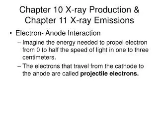
X-ray production • Push the “rotor” or “prep” button • Charges the filament – causes thermionic emission (e- cloud) • Begins rotating the anode. • Push the “exposure” or “x-ray” button • e-’s move toward anode target to produce x-rays HABIS
High Electrical Potential Electrons + - Radiation Penetrate the Sample Exposure Recording Device X-ray production (cont.) • The cathode contains a small filament much the same as in a light bulb. • Current is passed through the filament which heats it. The heat causes electrons to be stripped off. • The high voltage causes these “free” electrons to be pulled toward a target material (usually made of tungsten) located in the anode. • The electrons hit the target. This causes an energy exchange which causes x-rays to be created. filament HABIS
Interactions in the Body: • Three things can happen to x-rays as they hit the body: • Absorption (photoelectric effect) – x-ray is absorbed by tissues – does not contribute to image. • Scattered. • Transmission – penetrates through body to hit radiographic film or detectors. HABIS
Little electrons More electrons HABIS
Filtration • Beam filtration modifies the quantity and quality of the x-ray beam by removing low-energy(softer) photons in the spectrum. Three kinds of filtration: • Inherent – due to tube housing, insulation, etc. • Added – aluminum shielding that blocks low energy x-rays. ( 2 - 2.5mm) AL filter . • Special – used to image body parts that have varying thickness or density. (like wedge filters) • Filtration is measured in terms of “half-value layer” (HVL) HABIS
Collimation • Is located under the port of the X-ray tube. • Has a light in it for radiographer to see where x-rays would hit the patient. • Advantages: • ↓ patient dose • ↓ scatter radiation (↑contrast) • Always use the minimum acceptable field size • Collimation should be visible on a minimum of three sides of the film. HABIS
Effects of collimation (beam restriction) on scatter H kkuh
Contrast • ↑contrast = short scale = more black and white (less detail) • ↓contrast = long scale = mores shades of grey (more detail) • Controlling factor kVp • ↑kVp = ↓ contrast (more shades of grey) HABIS
Terms Related to Image Production • Attenuation • The process by which primary radiation is changed or absorbed as it travels through the patient • Radiolucent • Material that allow x-ray photons to pass through easily (air) • Radiopaque • Materials that do not allow x-ray photons to pass through easily (bone) can be seen on plain film. HABIS
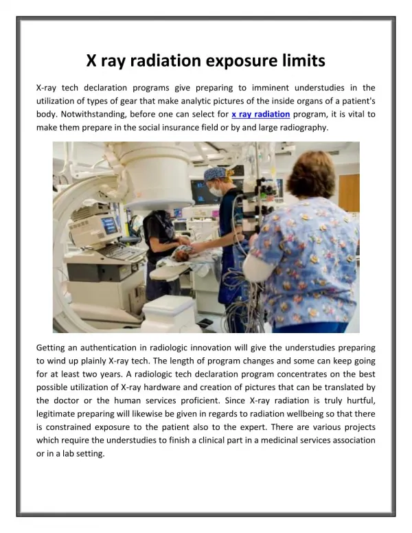
Reducing Exposure (protection) • Time Reduce the spent near the source of radiation. • Distance Increase the distance from the source of radiation. • Shielding Place shielding material between you and the source of radiation. HABIS
THINK ABOUT THIS H kkuh
THANK YOU NOTHING IMPOSIBLE HABIS