Download
1 / 40
400 likes | 536 Views
Central systems. Central systems – cells and circuits that mediate functions necessary for the coordinated behavior of the whole organism. The hypothalamus. The hypothalamus regulates basic life processes by three main mechanisms.

E N D
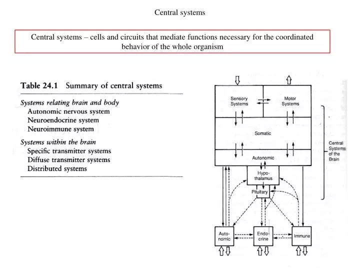
Central systems Central systems – cells and circuits that mediate functions necessary for the coordinated behavior of the whole organism
The hypothalamus • The hypothalamus regulates basic life processes by three main mechanisms. • First, the visual inputs are used by the suprachiasmatic nucleus to synchronize the internal clock mechanism to the day-night cycle in the external world. • Second, the hypothalamus compares sensory information with biological set points. There are set points for a wide variety of physiological processes, including blood sugar, sodium, osmolality, and hormone levels. • Finally, when the hypothalamus detects a deviation from a set point, it adjusts an array of autonomic, endocrine, and behavioral responses to restore homeostasis. E.g., if the body is too warm, the hypothalamus shifts blood flow from deep to cutaneous layers and increases sweating, to increase heat loss through the skin. Meanwhile, the hypothalamus activates coordinated behaviors, such as seeking to change the local ambient temperature or seeking a cooler environment.
Pituitary gland The master neurohemal organ (i.e., organ that releases stored neurosecretory substances into the blood ) in vertebrates is the pituitary gland. The hypothalamus controls the endocrine system through the pituitary directly and indirectly. The pituitary consists of an endocrine and a neural part. The endocrine (anterior) part is called adenohypophysis and neural (posterior) part is called neurohypophysis. Direct control: The neurohypophysics contains axons terminals (5) releasing two peptide hormones: oxytocin and vasopressin. Indirect control: Hypothalamic axons terminals (1-4) secrete regulatory hormones into the local circulation, which are carried by blood vessels into the anterior pituitary. These regulatory hormones control the synthesis and release of anterior pituitary hormones into the general circulation.
Hypothalamic – pituitary system Cells in different regions (hypothalamus and other sites) all send axons to the median eminence. Discharge of these neurons release so called releasing factors (RF). Their action on pituitary cells are to stimulate immediate release as well as to induce long term synthesis of hormones. Hypothalamus Pituitary
Neuroimmune circuits The main route of neural immunoregulation is the hypothalamic-pituitary-adrenal cortical axis (HPA) (oś podwzgórze – przysadka – kora nadnerczy). Within the immune system organs phagocytic (phag) and B and T lymphocytes proliferate in response to antigenic (Ag) stimulus. Low concentrations of corticosteroids (CS) and adrenocorticotropic hormone (ACTH) stimulate immunological response (+). High concentrations of CS and ACTH (thick arrows) suppresses immunological response (-). The substances of the immune system regulate the immune response but also act to block the actions of other substances at different levels of the HPA axis (black bars). The immunosupression in people suffering depression or stress is related to suppression of the immune response by CS and ACTH.
Neuroimmune circuits The time course of the antibody response of the immune system to an antigenic stimulus (above) and corticogenic response of HPA axis (below). There is clear correlation between the two responses. Between the graph, mechanism correlating the two systems are postulated.
Immune system – membrane properties A variety of substances can affect the immune response implying the presence of appropriate membrane receptors. Immune cells have a number of receptors which are involved in neural – immune interactions. A. Ligand-gated channels. B. Voltage-gated channels. Because of the many receptors present, some researche suggested that the certain cells of immune system may serve as „free-floating nerve cells”, and collectively may represent a mobile brain. It could serve as a sensory organ („sixth sense”) for stimuli such as bacteria, viruses and tumor cells.
Central systems Central systems – cells and circuits that mediate functions necessary for the coordinated behavior of the whole organism
Glutamate Distribution of neurons using glutamate as a neurotransmitter in the rat brain. Most prevalent neurotransmitter with a primarly excitatory action. Low and moderate glutamate levels, activate AMPA receptors High glutamate concentrations activate NMDA receptors Distribution of NMDA receptors in the rat brain. Highest receptor concentration include the olfactory bulb (OB), hippocampus (Hi), cerebellum (Cb). Intermediate receptor densities are seen in the cerebral cortex (Cx), striatum (St), thalamus (Th).
g-Aminobutyric Acid (GABA) Distribution of GABAergic neurons. There are short tracts from striatum to substantia nigra and in cerebellum. The long pathway projects form hypothalamus to cerebral cortex. Most GABAergic neurons are local interneurons in cortex, olfactory bulb, hippocampus, cerebellum and retina. Most prevalent neurotransmitter with a primarly inhibitory action. There are two classes of GABA receptors: GABAA and GABAB. There are numerous subunit isoforms for the GABAA receptor. The subunit composition determines which ligands may bind and modulate the receptors besides GABA itself. Examples: Barbiturates (hypnotics e.g. phenobarbital), Benzodiazepines (relaxants e.g. Valium), Ethanol (alcohol). Isoforms of GABAA receptors in different brain areas. Each region, subregion and layer has its unique combination of subunits.
Glycine Traditionally, glycine has been localized mainly to the brainstem and spinal cord. Recent years have brough the evidence of a widespread distribution of glycine in the brain. Glycine is an inhibitory neurotransmitter. Distribution of the b subunits of the glycine. High density of receptor is visible in the olfactory bulb (OB), hippocampus (Hi), cerebellum (Cb), cerebral cortex (Cx), striatum (St) and thalamus (Th). The detection of glycine in the interstellar medium has been debated. Kuan YJ, Charnley SB, Huang HC, et al., Interstellar glycine, Astrophys J (2003), 593(2): 848-867 „Glycine was probably formed when ices containing simple organic molecules were exposed to ultraviolet light” – if amino acids exsist in outer space than there might be proteins and life as well! Later, Kuan’s results were called into question: Snyder LE, Lovas FJ, Hollis JM, et al., A rigorous attempt to verify interstellar glycine, Astrophys J (2005), 619(2): 914-930. In 2008, the glycine-like molecule aminoacetonitrile was discovered in a giant gas cloud near the galactic center in the constellation Sagittarius by the Max Planck Institute for Radio Astronomy. In 2009, glycine sampled in 2004 from comet Wild 2 by the NASA spacecraft Stardust was confirmed.
Miller-Urey experiment (1953) Jupiter’s atmosphere – methane (CH4), ammonia (NH3), hydrogen (H2), helium (He). Simulations of the conditions of the early Earth’s atmosphere: water (H20), methane (CH4), ammonia (NH3), hydrogen (H2), carbon monoxide (CO). Experimental setup of Miller-Urey experiment - replication at NASA. At the end of two weeks of continuous operation 10–15% of the carbon within the system was in the form of organic compounds. Two percent of the carbon had formed amino acids that are used to make proteins in living cells, with glycine as the most abundant. Recent results: "old" genes, defined as those that are found to be common to organisms from several widely separated species are enriched in those amino acids that are also most readily produced in the Miller–Urey experiment. Miller S. L. (1953). "Production of Amino Acids Under Possible Primitive Earth Conditions". Science117: 528.
Dopamine • Loss of DA synapses in the striatum (in basal ganglia) is primary cause of movement disorders (e.g. Parkinson disease). • In hypothalamus DA is influences the release of the hormones, mainly prolactin. • In the limbic system DA is implicated in the reward system responsible for biological drives (feeding, drinking, sex) and motivation. Dopamine is important for immediate pleasant rewards and for arousal effects that are predictive of impending rewards. Distribution of dopamine-containing neurons. The two main projection neuron groups are located in substantia nigra (SN) and ventral tegmental area (VTA). Besides there are short axon DA cells in olfacory bulb, hypothalamus, retina. Main projections reach the limbic structures (septum, amygdala, prefrontal cortex) and are implicated in emotion and agression; prefrontal cortex is crucial for some of our highest cognitive functions.
Dopamine • Almost all addictive substances (eg. cocaine, opiates (morphine, heroin), nicotine or alcohol) increase the DA level in NA. • Addiction involves positive reinforcement related to increase of DA release and negative visceral side-effects related to drug withdrawal. • In people in the state of romantic love there is strong activation of dopaminergic pathways of the VTA; love is rather a motivation system than an emotion. Dopamine is also responsible for general arousal component for the romantic love. Brain-reward circuitry in the rat
Acetylocholine ACh acts on either nicotinic receptors (producing brief synaptic potentials) or muscarinic receptors (producing slow synaptic potentials). It is the only neurotransmitter used as an excitatory neurotransmitter at neuromuscular junctions in skeletal muscles. Acetylcholine is one of many neurotransmitters in the autonomic nervous system. In cardiac tissue acetylcholine has an inhibitory effect, which lowers heart rate. It produces both excitatory and inhibitory responses in the brain. ACh has an important role in the enhancement of sensory perceptions when we wake up and in sustaining attention. Damage to the cholinergic (acetylcholine-producing) system in the brain has been shown to be associated with dementia (Alzheimer’s disease). Distribution of cholinergic cell groups and their projections in the rat brain. Main projections are from habenula (HAB), septum (SEP), striatum (STR), basal nucleus (BN).
Norepinephrine The terms noradrenaline (from the Latin) and norepinephrine (from the Greek) are interchangeable. Norepinephrine (NE) or noradrenaline (NA) has neuromodulatory action and is setting levels of central neural activity for different behavioral states. It is released by Locus Ceruleus (LC) neurons to almost every regions of the CNS. NE neurons act like a central autonomic nervous system, to complement the peripheral autonomic system and its release of epinephrine (andrenaline) into the bloodstream. Distribution of norepinephrine containing neurons. The widespread patterns of projections suggest that these neurons are involved in adjustments of global neuronal excitability.
Serotonin Serotonin (5HT) is released by neurons in Raphe Nuclei, which project widely to many brain regions, alike the NE neurons. Like the NE system, the 5HT system exerts widespread influence over arousal, sensory perception, emotion and higher cognitive functions. Serotonin is thought to be a contributor to feelings of well-being and happiness. Several antidepressive drugs inhibit the reuptake of serotonin, making it stay in the synaptic cleft longer. The psychedelic drugs psilocin/psilocybin, DMT, mescaline, and LSD act as partial agonists at postsynaptic serotonergic receptors. They bind to 5HT receptors but their presence prevents serotonin from sending neural messages. It is redirected to other brain areas and causes psychedelic hallucinations. Distribution of neurons releasing serotonin. lysergic acid diethylamide serotonin
Peptides are short chains of amino acid monomers linked by peptide bonds. Polypeptides are long, continuous, and unbranched peptide chains. Proteins consist of one or more polypeptides arranged in a biologically functional way. Peptides - substance P Substance P is an important element in pain perception. The sensory function of substance P is thought to be related to the transmission of pain information into the CNS. Substance P has been associated with the regulation of mood disorders, anxiety, stress, reinforcement, neurogenesis, respiratory rhythm,neurotoxicity, nausea/vomiting. Distribution of substance P containing neurons.
Free nerve endings • The simplest type of sensory receptor in the skin is the free nerve ending. They respond to: • mechanical stimuli (mechanoreceptors) • heating and cooling (thermal receptors) • noxious stimuli (nociceptors – pain receptors) Activation of receptorsmediatingpain. Recording of the compoundactionpotential from a peripheralnerve in response to a single electricshock, showingdifferentcomponents of fast conducting (Aa, b), medium conducting (Ad) and slowconducting (C) fibers. The immediate sharppainismediated by Ad fibers, the subsequentconstantachingismediated by the C fibres.
Peptides - somatostatin Somatostatin regulates (inhibits) the secretion of growth hormone in anterior pituitary gland. Somatostatin is also secreted in several locations in the digestive system: stomach, intestine and pancreas. Distribution of somatostatin containing neurons. Somatostatin analogues are used to treat acromegaly.
Endorphin and enkephalin The short chain penta-peptides are called ekephalins whereas the longer chain compounds are called endorphins. The term endorphins may be used to refer to both. Endorphins ("endogenous morphine”) are produced by the pituitary gland and the hypothalamus during exercise, excitement, pain, consumption of spicy food, love and sex. They resemble the opiates in their abilities to produce analgesia and a feeling of well-being. Endorphins act on the μ1 opioid receptors, which are are presynaptic, and inhibit neurotransmitter release; through this mechanism, they inhibit the release of the inhibitory neurotransmitter GABA, and disinhibit the dopamine pathways, causing more dopamine to be released. Opioids are among the world's oldest known drugs (opium). Distribution of endorphin containing neurons. Capsaicin is the chemical which give rise to the heat of chillies. When eaten, it stimulates body's production of endorphins.
Emotion and feeling “The value of life can be measured by how many times your soul has been deeply stirred.” Soichiro Honda
The beginnings of studies of emotions Darwin’s dog, displaying the contrasting expressions of hostility and affection. From: The Expression of Emotions in Man and Animals, Charles Darwin. Darwin first pointed out in 1872, that fearful, angry, and happy facial expressions are universal. The Expression of Emotions in Man and Animals, Charles Darwin, 1872
Emotion and feeling An emotional state has two components, the one is a characteristic physical sensation and the other is a conscious feeling. The term emotion sometimes is used to refer only to the bodily state (i.e., the emotional state) and the term feeling is used to refer to conscious sensation. Conscious feeling is mediated by the cerebral cortex, in part by the cingulate cortex and by the frontal lobes. Emotional states are mediated by a family of peripheral, autonomic, endocrine, and skeletomotor responses. These responses involve subcortical structures: the amygdala, the hypothalamus, and the brain stem. stimulus Model of the basic neural systems that control emotions. The particular emotion experienced is a function of cross-talk between neocortical and subcortical structures, as well as feedback from peripheral receptors.
First theories of emotions James-Lange: Emotions are cortical interpretation of feedback information from the periphery. Cannon-Bard: Emotional experience and physiological arousal are separate and are both triggered by subcortical activity. Traditionally, it was assumed that emotions are initiated by cognitive activity. James – Lange (1884) proposed the opposite view, that the cognitive experience of emotion is secondary to the physiological expression of emotion. He suggested that when we encounter a potentially dangerous situation, we act instinctively by running away and then consciously explain our action and the changes in our body (the increase in heart rate and respiration) as “driven by fear.” Experimental support for the James-Lange theory exists. 1) objectively distinguishable emotions can be correlated with specific patterns of autonomic, endocrine, and voluntary responses. 2) patients with the spinal cord transection, whom lack feedback from the autonomic nervous system appear to experience a reduction in the intensity of their emotions. The James-Lange theory also have shortcomings: for example, one often continues to be emotionally aroused even after the physiological changes have subsided. According to Cannon and Bard (1915), the physiological responses to emotionally significant stimuli are too undifferentiated to convey to the cortex specific, detailed information about the nature of an emotional event. They showed that intense emotion triggered an emergency reaction—a fight-or-flight response —in anticipation of additional behavioral responses and expenditure of energy. Cannon suggested that this flight-or-fight response was mediated by the sympathetic component of the autonomic nervous system and that it acted as a whole, almost in an all-or-none way, independent of the specific emotionally significant stimuli that elicited it. They proposed that perceived stimulus (by the thalamus) triggers both physiological response (mediated by the hypothalamus) and emotional experience (mediated by the cortex); physiological arousal does not have to precede emotional experience.
The Schachter’s theory Schachter proposed (1960s) that the cortex actively translates peripheral signals into specific feelings. He suggested that the cortex creates a cognitive response to ambiguous peripheral information consistent with the individual's expectations and social context. Experimental support: Schachter injected volunteers with epinephrine; some subjects were informed of the side effects (eg, pounding heart), others were not. All of the subjects were then exposed either to annoying or amusing conditions. The subjects who had been warned about the side effects of epinephrine exhibited less anger or less pleasurable feelings.
The Arnold’s theory According to Arnold (1950 - 1970) an emotion is the product of unconscious evaluation of a situation as potentially harmful or beneficial, while feeling is the conscious reflection of the unconscious appraisal. Feeling is therefore a tendency to respond in a particular way, not the response itself. Emotions are different because they elicit different action tendencies. The appraisal theory accounts for individual variances of emotional reactions to the same event. A finding supporting this idea is that we can have emotional reactions to subliminal stimuli. Unlike the James-Lange theory, Arnold's view does not require that we have an autonomic response to experience emotion.
Hypothalamic mechanisms Walter Cannon and Philp Bard examined behavior of cats after transections of the brain at different level. Electrode (A) was used to stimulated the hypothalamus and produce expressions of rage and fear. Transection (B) removing the forebrain and leaving the hypothalamus produces sham rage (furia pozorna), which lacks conscious manifestations of normal attack behavior. After transection (C) below the hypothalamus the sham rage response is lost.
Agressive behavior Selective stimulation of sites in the hypothalamus may induce two basic types of attack. In affective attack (above) the animal displays sympathetic arousal, emotional exciment and rage. In quiet biting (below) the cat shows no emotion but proceeds to caputre the rat and bite it.
Papez circuit for emotions According to Papez (1937) neural circuit underying emotions involves the limbic system. It starts with the mammilary body of the hypothalamus as the site of output for expression of the emotions (1). Collateral fibers pass to the thalamus (2) and cingulate gyrus (3). Here, it was proposed that conscious, subjective emotional experience arises. The cingulate gyrus project to the hippocampus (4). The hippocampus integrates different inputs and projects them to the mammilary body (5). A neural circuit for emotion extended by Paul MacLean. The circuit originally proposed by Papez is indicated by thick lines; more recently described connections are shown by fine lines. Known projections of the fornix to hypothalamic regions (mammillary bodies and other hypothalamic areas) and of the hypothalamus to the prefrontal cortex are indicated. A pathway interconnecting the amygdala to the hypothalamus is shown. Finally, reciprocal connections between the hippocampal formation and the association cortex are indicated.
The amygdala is the part of the limbic system most specifically involved with emotional experience The amygdala Brain imaging studies (MRI + PET + fMRI) demonstrate the role of the amygdala in emotional responses A. A series of faces shows a continuum of expression between happiness and fear. Activity in the brain of normal subjects was recorded as they viewed these faces. B. With the presentation of each of the faces only the left amygdala was found to vary in a systematic fashion. C. The mean regional cerebral blood flow (rCBF = PET or fMRI) shows that the responses in the amygdala were significantly greater to fearful expressions than to happy expressions. These results are consistent with recording and ablation experiments on animals that suggest the amygdala has a critical role in emotions, particularly in fear.
Fear conditioning The amygdala mediates both inborn and acquired emotional responses. The best studied example of a learned emotional state is the classical conditioning of fear. Bilateral lesions of the basolateral complex of the amygdala in experimental animals abolish this learned response to fear. In this form of learning an initially neutral stimulus, such as a sound that does not evoke autonomic responses, is paired with an electric shock to the feet, which produces pain, fear, and autonomic responses. After several pairings the sound itself elicits a fearful reaction, such as freezing in place or changes in heart rate or blood pressure.
The amygdala The amygdala appears to be involved in mediating both the unconscious emotional state and conscious feeling. Consistent with this dual function of emotion, the amygdala has two projections. Many of the autonomic expressions of emotional states are mediated by the amygdala through its connections to the hypothalamus and the autonomic nervous system. The influence of the amygdala on conscious feeling is mediated by its projections to the cingulate gyrus and prefrontal cortex. Electrical stimulation of the central nucleus of the amygdala produces a variety of effects
Facial expression - muscles Dermal muscles of the human face as portrayed in Darwina’s book (1872). In higher mammals and especially in humans facial muscles have become adapted for the expression of emotions.
Pathways for facial muscles control Have a look at: http://www.artnatomia.net/uk/artnatomy2014.html Neural circuits mediating control of the dermal muscles of the face. Social (Pan-Am) smile Spontaneous smile
Facial expression - evolution Comparison of human expression of smiling and laughter with bared-teeth displays of lower primates and primitive mammals. Friendly human smile evolved from more aggressive display of teeth.
The prefrontal, cingulate and parahippocampal cortices ACG – cingulate gyrus OFC – orbitofrontal cortex PFC – prefrontal cortex Cingulate gyrus has numerous connections to a variety of cortical and subcortical structures. It may act as a control center between these structures. Cortical mechanisms provide a means by which memory and imagination, not just external stimuli, can evoke emotional feelings and they enable us to use emotional information generally in cognitive processing. Cortical structures also provide the means by which conscious thought can suppress reflex emotional responses.
The frontal cortices - Phineas Gage and hazard The executive functions of behavior—judgment, long-term planning, and holding and organizing events from memory for prospective action take place in the anterior association area (prefrontal cortex). The ventromedial frontal cortex is thought to provide source of cognitive control of emotional responses by predicting their consequences. Lesions to the ventral sector of the frontal lobe result in disinhibition of inappropriate behavior in social situations. A B C D +100 +100 +50 +50 -1250 -1250 -100 -100 In a “gambling experiment” a player sits draws cards from four decks of cards, labeled A, B, C, and D. He is given a loan of $2000. The undisclosed rules are that A and B cards yield $100 but occasionally require the subject to repay $1250. Cards C and D yield $50 but only require repayment of small sums (less than $100). Normal people initially play decks A and B, but gradually, usually within 30 of the 100 trials switch to a preference for decks C and D. Patients with frontal lesions prefer cards from decks A and B throughout the test despite the high penalties and the need to borrow from the bank. Insights into the function of cortical association areas has come from the case of Phineas Gage. An iron bar was driven through his frontal lobes by an explosion. His personality was remarkably changed after the accident. Before the accident he had been reliable and calm but afterward he became unreliable and often drank excessively. He was unable to manage his work or personal life, and eventually became unemployed and homeless.
Effect of meditation on brain function and structure Meditation increases activation and grey matter concentration in brain regions involved in learning and memory, emotion regulation, sense of self. Frontal cortical areas are thicker in meditators. Lazar et al., Meditation experience is associated with increased cortical thickness. Neuroreport. 2005; 16(17): 1893–1897. Stronger brain fMRI activation in the anterior cingulate cortex and medial prefrontal cortex in meditators than controls for the contrast mindfulness>arithmetic. Hoelzel et al., Differential engagement of anterior cingulate and adjacent medial frontal cortex in adept meditators and non-meditators. Neuroscience Letters 421 (2007) 16–21