Download
1 / 30
300 likes | 489 Views
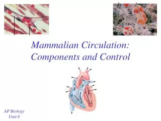
Mammalian Circulation: Components and Control. AP Biology Unit 6. Mammalian Heart. Right side. Left side. 4 chambered heart (2 atria, 2 ventricles) Valves = flaps that keep chambers of the heart closed at the right time

E N D
Mammalian Circulation: Components and Control AP Biology Unit 6
Mammalian Heart Right side Left side • 4 chambered heart (2 atria, 2 ventricles) • Valves = flaps that keep chambers of the heart closed at the right time • Valves are needed to build pressure in heart and prevent back-flow of blood.
Atrioventricular (AV) Valves • Located between the atria and ventricles • Tricuspid Valve • Between the right atrium and right ventricle • 3 flaps • Bicuspid (Mitral) valve • Between the left atrium and left ventricle • 2 flaps
Semilunar valves • located at two exits for the heart • Between the right ventricle and the pulmonary artery (to lungs) • Between the left ventricle and the aorta (to the body)
Pathway of Blood • Do you remember the pathway of blood through the body and the heart? • Use these terms: Right Atrium, Left Atrium, Right Ventricle, Left Ventricle, Pulmonary Artery, Pulmonary Vein, Aorta, Lung Capillaries, Capillaries in Top or Bottom of Body, Anterior / Posterior Vena Cava • Start where the blood first leaves the heart to go to the body
Pathway of blood • Aorta arteries capillaries in body veins vena cava right atrium right ventricle pulmonary artery lung capillaries pulmonary vein left atrium left ventricle aorta
Questions… • Where does the blood have the highest O2 concentration? • Just after leaving lungs (where it picked up O2) • Where does the blood have the highest CO2 concentration? • Just before getting to the lungs (hasn’t dropped off the CO2 waste yet)
Heartbeat • The heart beat is controlled by electrical signals generated in specific cells in the heart = self excitation • Sinoatrial (SA) node = a group of specialized cells that initiates the heartbeat • Also called the pacemaker of the heart • generates electrical impulses that cause both atria to contract
Heartbeat • Atrioventricular (AV) node • When it receives the signals from the SA node, it transfers the signals to the Bundle of His • Bundle of His spreads the signal to the Purkinje fibers in the ventricles both ventricles contract
Blood Flow through Vessels • Arteries (and arterioles) • Thick, muscular walls • Walls also contain collagen and elastic fibers (make them stretchable) • Can be constricted or dilated
Blood Flow through Vessels • Veins (and venules) • Thinner walls • one-way valves to prevent blood from flowing backwards • Blood in veins is moved by the contractions of skeletal muscles around them (“milking”) – low pressure
Blood Flow through Vessels • Capillaries • Thin walls (usually one cell layer thick) • Permeable to water, ions, other small molecules • Blood flows slowly through them (red blood cells often have to travel single file) • Every cell in the body is close to at least 1 capillary
Capillaries • Capillaries exchange materials between the blood and the interstitial fluid • Blood pressure and osmotic pressure drive the movement of molecules into and out of capillaries • Blood pressure forces water and solutes out (on the artery side) • Osmotic pressure (due to the proteins left in the capillaries) causes fluid to flow back into the capillaries by osmosis
Question… • How does the structure of each type of vessel support its function? • Arteries– thick walls can withstand pressure from heart pumping blood • Veins- valves help prevent backflow since the heart is too far away to provide forward pressure • Capillaries- very thin walls allow for easy exchange with the interstitial fluid
Blood Pressure • Blood pressure = the force being applied to the blood vessel walls (from blood). • 2 phases of the cardiac cycle • Systole = when the heart muscle is contracting • Diastole = when the heart muscle is relaxed (between contractions)
Blood Pressure • Systolic Pressure = pressure in arteries when heart contracts • Diastolic Pressure = pressure in arteries between contractions
Question… • Giraffes need higher blood pressure. Why? • Since they are taller, they need more pressure to get the blood all the way to the top of their bodies
Components of blood • Plasma (liquid) • Water, nutrients, proteins, ions, etc. • Cellular Components • Red blood cells (carry oxygen) • White blood cells (immune function) • Platelets (clotting)
Differentiation of Blood Cells • Blood cells (RBC, WBC and platelets) all develop from stem cells in the red bone marrow. • Erythropoietin (EPO) is a hormone that promotes the production of erythrocytes (RBC) • Synthesized in the kidneys
Question… • When the body is not receiving enough O2, what will happen to EPO levels? • They increase to create more RBC to carry O2
Control of Circulation • Heart rate is controlled by • Nerve impulses sent to SA and AV Nodes • Parasympathetic division- slows heartbeat down • Sympathetic division – speeds up heart beat • Hormones (adrenaline/epinephrine) • Body temperature • Oxygen requirements due to exercise
Control of circulation • The opening of sphincters leading to capillary beds (group of capillaries) is controlled by • Nerve impulses • Hormones • Allows blood to be directed to specific parts of the body under stressful conditions
Control of Circulation • The Lymphatic System also plays a role in controlling circulation • Lymph = fluid in lymphatic system (like interstitial fluid, high in water and other nutrients) • Fluids flow out and into lymph capillaries via blood pressure and osmotic pressure • Maintains blood volume so blood pressure can remain constant
Question… • How is the lymph connected to the digestive system? • Lacteals are lymph vessels in the villi that absorb some nutrients from the small intestines.
Blood clotting • Platelets begin the clotting reaction • damage in the blood vessel wall exposed collagen fibers • Platelets stick the collagen release substances to make other platelets sticky • Clotting factors = released by platelets to activate enzymes needed for clotting
Blood Clotting STEPS: • Platelets adhere to the damaged region and become sticky release clotting factors • Clotting factors cause Prothrombin (inactive) to become Thrombin (active) • Thrombin causes Fibrinogen (plasma protein) to become fibrin (active form) • Fibrin forms threads that help seal the damaged area up
Cardiovascular Disease • Atherosclerosis = narrowing of arteries due to plaque build up • plaque deposits as a result damage to the vessel lining • Plaque deposits narrow the pathway for blood to flow • If the plaque is ruptured it will also cause clotting to occur– blocks pathway of blood
Cardiovascular Disease • Atherosclerosis can lead to heart attack or stroke • Heart attack – blockage in the arteries that supply the heart with blood • Stroke = blockage in an artery in the brain • Relationship between high blood pressure and heart disease? • High blood pressure will damage the lining of the arteries causes plaque to deposit in the damaged areas.
Heart Disease and Cholesterol • Cholesterol travels in the blood (plasma); carried by lipoproteins • Low Density Lipoproteins (LDLs) are associated with cholesterol deposits in arteries = “bad” cholesterol • High Density Lipoproteins (HDLs) appear to reduce cholesterol deposition = “good” cholesterol • What seems to matter is the ratio of HDL to LDLs in your blood
Cholesterol and lifestyle choices • While there is a genetic component to cholesterol levels, lifestyle choices also influence it • Exercise increases HDL levels, lowers LDL levels • Smoking increases LDL and lowers HDL levels