Download
1 / 56
560 likes | 765 Views
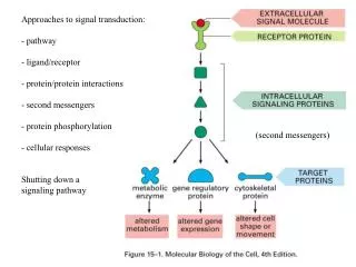
Biosensors to detect enzyme-ligand and Protein-protein interactions . -Physical parameters in binding studies-principles, techniques and instrumentation -Methods to probe non-covalent macromolecular interaction (stopped-flow, BIAcore, and Microcalorimetry)

E N D
Biosensors to detect enzyme-ligand and Protein-protein interactions -Physical parameters in binding studies-principles, techniques and instrumentation -Methods to probe non-covalent macromolecular interaction (stopped-flow, BIAcore, and Microcalorimetry) Lecturer: Po-Huang Liang 梁博煌, Associate Research Fellow Institute of Biological Chemistry, Academia Sinica Tel: 27855696 ext. 6070
Stopped-flow for measurements of protein-protein and protein-small molecule interaction
Substrate binding kinetics k1[S] Rate = d[E]/dt = -k1[S][E] d[E]/[E] = -k1[S]dt ln([E]t / [E]o) = -k1[S]t [E]t = [E]o exp (-k1[S]t) [ES] = [E]o-[E]t = [E]o(1-exp (-k1[S]t)) kobs = k1 [S] E ES k1[S] kobs = k1[S] + k-1 The slope of kobs vs [S] gives kon and intercept gives koff E ES k-1
3-D structure of E. coli UPPs Two conformers were found: one (closed form) with Triton bound and the other (open form) has empty active site Ko, T. P. et al, (2001) J. Biol. Chem 276,47474-47482.
Substrate-binding site The amino acids in a1 area are important for catalysis and substrate binding D26 is located in a P-loop conserved for pyrophosphate binding Pan et al., (2000) Biochemistry 39, 13856-13861
Large L137 on the bottom controls product chain length upper: + Triton bottom: no Triton
Long-lived intermediate C30 formed by A69L and C35 by L67W A69L L67W
Synthesis of FsPP to Probe UPPs Conformational Change Chen et al.(2002) J. Biol. Chem. 277, 7369-7376. FsPP Ki of FsPP as an inhibitor = 0.2 mM kcat of FsPP as an alternative substrate = 3 x 10⁻⁷ s-1
Stopped-Flow experiments UPPs-FPP + IPP UPPs-FsPP + IPP 3 phases in 10 sec 1 phase in 0.2 sec Binding rates vs. [IPP] gives IPP kon = 2 mM-1 s-1 2 phases in 0.2 sec
Different level of Trp fluorescence quench by FPP wild-type W31F has less quench FPP binding mainly quenches the fluorescence of W91, a residue in the a3 helix that moves toward the active site during substrate binding W91F has almost no quench
FPP binding does not require Mg2+; IPP binding needs Mg2+ FPP (or FsPP) quenches the UPPs Intrinsic fluorescence even in the absence of Mg2+ Mg2+ is required fro IPP binding + Mg2+
The role of a flexible loop of residues 71-83 The invisible loop in the E. coli UPPs structure is responsible to bring IPP to the correct position and orientation to react with FPP Ko et al., (2001) J. Biol. Chem. 47474-47482
Fluorescent probe for ligand interaction and inhibitor binding Chen et al., (2002) J. Am. Chem. Soc. In press
Characterization of the fluorescent probe (A) (B) (A) Fluorescence is quenched by UPPs and recovered by replacement with FPP (B) Probe binds to UPPs with 1:1 stoichiometry (C) (D) (C ) Probe binds to UPPs with a kon = 75 mM-1 s-1 (D) Probe releases from UPPs (chased by FPP) with a koff = 31 s-1
Substrate and product release rate FPP is released at 30 s-1 UPP is released at 0.5 s-1 Can this method apply to drug-targeted prenyltransferases to find non-competitive inhibitor?
Reaction: DHF + NADPH THF + NADP+ Association:
Competition experiments to measure Dissociation rate constants using Stopped-flow
Rate constant for the pre-steady-state burst measured by stopped-flow energy transfer Uisng NADPH, 450 s-1 is followed by a 12 s-1 steady-state rate. Using NADPD, 150 s-1 is followed by the same rate at pH 6.5, isotope effect kH/kD =3 Pre-steady-state rate is decreased with pH
Observed rate constants for hydride transfer as a function of pH and predictable kinetic behavior
Interaction of colicin E7 and immunity 7 Entrance of colicin E is through the BtuB (vitamin B12 receptor) and TolA
Wallis et al., Protein-protein interactions in colicin E9 Dnase – immunity protein complexes. 1. Diffusion controlled association binding for the cognate complex Biochemistry 1995, 34, 13743-13750 Association kinetics of ColE9-Im9 complex (lower) and E9-DNase-Im9 (upper), [ColE9] = 0.35 mM, [Im] = 1.75 – 7 mM. Dissociation kinetics of ColE9-Im9 complex at 0 and 200 mM NaCl. The preincubated E9 with [3H]Im (6 mM) was mixed with unlabeled Im (54 mM) k2 k1 kon = 4 x 10⁹ M-1 s-1 koff = 3.7 x 10⁻⁷ s-1 Kd = koff /kon = 7.2 x 10⁻¹⁷ M N + I NI* NI k-1 K-2
Sensor chip and coupling CM5: couple ligand covalently NTA: bind His-tagged lignad SA: capture biotinylated biomolecules HPA: anchor membrane bound ligand
Objects of the experiments • Yes/No binding, ligand fishing • Kinetic rate analysis ka, kd • Equilibrium analysis, KA, KD • Concentration analysis, active concentration, • solution equilibrium, inhibition Control of flow rate (ml/min) and immobilized level (RU) for different experiments
Definition ka A + B AB kd • Association rate constant: ka (M-1 s-1) • ---Range: 103 to 107 • ---called kon, k1 • Dissociation rate constant: kd (s-1) • ---Range: 10-5 to 10-2 • ---called koff, k-1 • Equilibrium constant: KA (M-1), KD (M) • ---KA = ka/kd = [AB]/[A][B] • ---KD = kd/ka = [A][B]/[AB] • ---range: pm to uM
Association and dissociation rate constant measurements ka A + B AB kd In solution at any time t : [A]t = [A]o – [AB]; [B]t = [B]o – [AB] d[AB]/dt = ka[A]t[B]t – kd[AB]t In BIAcore at any time t: [A]t = C; [AB] = R; [B]o = Rmax thus [B]t = Rmax – R d[R]/dt = ka*C*(Rmax-Rt) – kd (R)
It is easy to mis-interpret the data It Distinguish between fast binding and bulk effect: use reference or double reference Two ways to overcome mass transfer limitation: 1.increase flow rate 2. reduce ligand density
Example 2: Lackmann et al., (1996) Purification of a ligand for the EPH-like receptor using a biosensor-based affinity detection approach. PNAS 93, 2523 (ligand fishing) • Phenyl-Sepharose • Q-Sepharose HEK affinity column
Ion-exchange RP-HPLC
The ligand is Al-1, which is previous found as ligand for EPH-like RTK family
BIAcore analysis of bovine Insulin-like Growth Factor (IGF)-binding protein-2 Identifies major IGF binding site determination in both the N- and C-terminal domains J. Biol. Chem. (2001) 276, 27120-27128. IGFBPs contain Cys-rich N- and C-terminal and a linker domains. The truncated bIGFBP-2 were generated and their interaction with IGF were studied.
Lane 2: 1-279 IGFBP-2His Lane 3: 1-132 IGFBP-2 Lane 4: 1-185 IGFBP-2 Lane 5: 96-279 IGFBP-2His Lane 6: 136-279 IGFBP-2His
MicroCalorimetry System Right: ITC (Isotheromal titration Calorimetry) Inject “ligand” into “macromolecule” A small constant power is applied to the reference To make DT1 (Ts – Tr) negative. A cell feed-back (CFB) supplies power on a heater on the sample cell to drives the DT1 back to zero.
Binding isotherms Simulated isotherms for different c values c = K (binding constant) x macromolecule concentration c should be between 1 and 1000 Make 10-20 injections can be used to obtain binding affinity or binding equilibrium constant (Keq), molecular ration or binding stoichiometry (n), And heat or enthalpy (DH).
Activation of Ras following binding of a hormone (e.g. EGF) to an RTK GRB2 binds to a specific phosphotyrosine on the activated RTK and to Sos, which in turn reacts with inactive Ras-GDP. The GEF activity of Sos then promotes the formation of the active Ras-GTP.
Example: O’Brien et al., Alternative modes of binding of proteins with tandem SH2 domains (2000) Protein Sci. 9, 570-579 • pY110/112 bisphosphopeptide • binds to ZAP70 showing a 1:1 complex • (B) Monophoshorylated pY740 binds • to p85 with two binding events • (C) Binding of pY740/751 peptide into • p85. The asymmetry of the isotherm • shows two distinct binding events • showing that an initial 2:1 complex of • protein to peptide is formed. As further • peptide is titrated, a 1:1 complex is • formed.
ITC data for the binding of peptides to ZAP70, p85, NiC, and isolated c-SH2 domain KB1 and KB2 correspond to the equilibrium binding constants for the first and the second binding events.
Conformational change of two SH2 binding with phosphorylated peptide (A) Primary sequence NiC (B) a. NiC; b.NiC + bisphosphorylated peptide (C ) a. N-terminal SH2 alone; b.N-terminal SH2 + pY751 peptide; c. C-terminal SH2; .d. C-terminal SH2 + pY751 peptide