Download

1 / 26
340 likes | 791 Views
Anatomy& innervations of parotid,Submandibular &Sublingual Glands. parotid gland. It is the largest salivary gland (serous). It is located in a deep space behind ramus of mandible & in front of sternocleidomastoid.

E N D
Anatomy& innervations of parotid,Submandibular &Sublingual Glands
parotid gland • It is the largest salivarygland (serous). • It is located in a deep space behindramus of mandible & in front of sternocleidomastoid. • It is wedge shaped , with its base(concave upper end) lies above and related to cartilaginous part of external acoustic meatus/ and its apex (lower end) lies below & behind angle of mandible. • It has 2 borders :anterior convex border + straight posterior border. • Facial N. passes within the gland and divides it into superficial& deepparts or lobes.
Processes of the parotid gland : • It has 4 processes. • Superior margin of the gland extends upward behindtemporo-mandibular joint into mandibularfossa of skull ….. Glenoid process. • Anterior margin of the gland extends forward superficial tomasseter…facial process. • A small part of facial process may be separate from main gland…accessory part of gland,thatlies superficial to masseter. • Deep part of gland may extend between medial pterygoid & ramus of mandible …pterygoid process.
Capsules of the Gland & Parotid Duct : • It is surrounded by2 capsules,the first is C.T. capsule,the second is the dense fascial capsule of investing layer of deep cervicalfascia, (part of it is thickened to form stylomandibular ligament). • Parotid duct 5 cm.long, passes from anterior border of gland , superficial to masseter one fingerbreadth, below zygomatic arch, then it pierces buccal pad of fat & buccinator muscle. • It passes obliquley betweenbuccinator & m.m.of mouth (serves as valvelike mechanism to prevent inflation of duct during violent blowing) and finally opens into vestibule of mouth ,opposite upper 2nd molar tooth
Structures within the parotid gland From lateral to medial (horizontal section) : 1-Facial N.---- emerges from stylomastoid foramento enter the gland at its posteromedial surface,and divides into 5 terminal branches. 2-Retromandibular vein: is formed within the gland by Union of superf. temporal + maxillary veins. 3-External carotid artery and its 2-terminal branches 4-parotid group of lymph nodes.
Structures within the parotid gland 1-Facial N.----divides into 5 terminal branches, whichleaveanteromedial surface of the gland. 2-Retromandibular vein:it leaves lower end of the gland. It divides into anterior & posterior divisions. The ant.division joins facial v., / and the post. division joins the post.auricular v. to form ext.J.V. 3-External carotid artery : it divides into superficial temporal & maxillary arteries at neck of mandible, which they leave upper end & anteriomedial surface of gland.
Structures which enter or leave the gland • Upper end :enter : superficial temporal vein. Leave :1-superficial temporal artery. 2-auriculotemporal N. 3-temporal branch of facial N. • Lower end :leave :1-cervical branch of facial N. 2-retromandibular (posterior facial) vein & its 2 division. • Posteromedial surface :enter :1-external carotid artery. 2-facial N. • Anteromedial surface :leave : maxillary artery. enter : maxillary vein. • Anterior border :leave :1-zygomatic branch of facial N. 2-buccal branch of facial N. 3-mandibular branch of facial N. 4-parotid duct. 5- transverse facial artery (branch of ext. carotid artery)
Relations of the parotid gland Superficial R.(lateral R.) : Skin, + superficial fascia cotaining platysma & great auricular N. + deep fascia (parotid fascial capsule) + parotid L.Ns. Superior R. :ext.auditory meatus & posterior surface of temporo-mandibular joint. Antero-medial R. : ramus of mandible + temporomandibular j.+ medial pterygoid + masseter. Postero-medial R.:mastoid process & attached Ms. + styloid process & its attached Ms. + carotidsheath & its contents + Facial N. enters gland from its postero-medial surface + externalcarotid artery grooves gland at its posteromedial surface, then passes inside it.
Blood Supply, lymph drainage, and Nerve supply of parotid gland • Blood supply:external carotid A.+its 2-terminal branches (maxillary Ar. + superficial temporal Ar.)& The 2-veins (maxillary & superficial temporal veins) drain into the retromandibular vein. • Lymph D.:parotid L. Ns., which finally drain into deep C.L.Ns. • N.supply:1-Parasymp.secretomotor Fs.:Inferior salivary nucleus of 9th C.N. in M.O.—Viaits Tympanic branch to form tympanic plexus in middle ear, and then Via Lesser petrosal N.(preganglionic fibres) — into OTIC ganglion. Postgang. Secretomotor parasymp. Fs. : from otic ganglion Viaauriculotemporal .N. to supplyparotid gland. 2-Sympath.Fs. : plexus of nerves around external carotid artery. 3-Sensory Fs. :auriculotemporal N. (branch of mandibular N.), ascends from upper end of parotid gland to supply skin of scalp above auricle + great auricular N.(C2,3)
Parasympathetic secretomotor + Sensory Supply of parotid gland : • Otic ganglion is a small parasympathetic ganglion that is functionally associated with glossopharyngeal N. it is located in the infratemporal fossa, just below foramen ovale, medial to mandibular N. -Auriculo-temporal N. is a branch of post. division of mandibular N. + Great auricular N. (C2,3 ).
Clinical notes: 1-parotid gland infection---MumpsGland becomes swollen,painful because fascial capsule derived frominvesting layer of deep cervical fascia is strong and limits the swelling of gland. 2-Frey’s Syndrome : -it is an intersting complication that sometimes occurs after penetratingwounds of parotid gland. -when patient eats, beads of perspiration appear on the skin of parotid. -It is caused by damage to auriculotemporal & great auricular nerves. -During healing, parasymp.secretory Fs. in auriculotemporal N.grow out and join distal end ofgreat auricular N.(C2,3) supplying skin overparotid. These fibres reach the sweat glands in skin of face so, there is sweating on skin covering parotid, instead of salivation during eating.
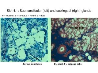
II. Submandibular salivary gland. • It is a lobulated mass, composed of serous & mucous acini. • It is surrounded by C-T capsule + densefascial capsule derived from investing layer of deep cervical fascia. • It has a large superficial part & a smalldeep part. • Its deep part is continuous with superficial part around posterior border of mylohyoid muscle. • Its superficial part lies in digastric triangle between mylohyoid & body of mandible (superficial to mylohyoid). • Its small deep part lies deep tomylohyoid and superficial to hyoglossus. Lateral view of submandibular & sublingual glands.
Relations of superficial part of Submandibular salivary gland. • Anteriorly : anterior belly of digastric • Posteriorly : posterior belly of digastric + stylohyoid muscle. • Medially (deep) :-mylohyoid.
Relations of superficial part of submandibular salivary gland. • Laterally : -it lies in contact with submandibular fossaon medial surface of mandible. • Inferolaterally (superficial) :-skin,superficial fascia, platysma & investing layer of deep cervical fascia + submandibular L.Ns. -it is crossed by facial vein & cervical branch of facial nerve.-facial artery ascends into digastric triangle, it deeply grooves posterior endof the gland, then passes betweenlateral surface of gland & the boneto reachbase of mandible where it pierces deep fascia to ascend to face.
Relations of Deep part of submandibular gland. • Medially (deep) : hyoglossus & styloglossus. • Laterally (superficial) : mylohyoid & superficial part of gland. • Superiorly : lingual N. & submandibular ganglion. • Inferiorly : hypoglossal N.
Submandibular Duct • It emerges from anterior end of its deep part. • It passes beneath m.m.of floor of mouth. • It is crossed by lingual N. ,then lies between sublingual gland & genioglossus muscle. • It opens into floor of mouth at the side of frenulum of tongue.
Note, frenulum of the tongue in midline = it is a fold ofm.m. connects undersurface of tongue to the floor of mouth. • Note, opening of submandibular duct into floor of mouth at the side of frenulum of tongue.
Bl. Supply : branches of Facial & lingual arteries. • Lymph drainage : submandibular + deep cervical L.N. • Nerve supply :1-Parasym.secretomotor Fs. :. from sup.salivary.N.of 7th C.N. (Facial N.) via chorda tympani N. to join lingual N. and pass into submandibular ganglion, then postganglionic parasymp. secretory Fs. From ganglion via lingual N. into gland. 2-Symp.Fs. : fromplexus of nerves around Facial +Lingual arteries. 3-Sensory :lingual N.
Clinical Notes • Calculus formation : its commmon site is the submandibular gland : tense swelling below the body of the mandible,which is greatestduring a meal and is reduced in size or absentbetween meals (diagnostic of the case). Clinically: by examination of floor of mouth, reveals absence of ejection of saliva from the orifice of duct.+ stone can be palpated in theduct,which lies below m.m. of the floor of mouth.
Clinical Notes : • Enlargement of Submandibular Lymph Nodesare commonly due to :1-Pathologic condition of scalp, face,maxillary sinus, or mouth cavity.2-Acute infection of teeth (most common cause of painful enlargement these nodes)
III. Sublingual salivary gl. • It is the smallest of the three main salivary glands. • It contains both serous & mucous acini. • It lies beneath m.m.of floor of mouth, within sublingual fold close to midline. • Sublingual ducts :The gland opens by numerous small ducts into floor of mouth on the summit of sublingual fold.
Note, openings of sublingual ducts 8-20 in number, they open into the floor of mouth on the summit of sublingual fold.
Relations of Sublingual salivary gland • Posteriorly : deep part of submandibular gland. • Medially (deep) : genioglossus +lingual N. + submandibular duct. • Laterally (superficial) :sublingual fossa of medial surface of mandible. • Superiorly :m.m. of floor ofmouth, forming sublingual fold. • Inferiorly : it is supported by mylohyoid muscle.
Blood supply, lymph drainage, ----- As the submandibular gland. • Nerve supply : 1- Parasymp.secretomotor Fs.—as submand.gl. and postganglionic Fs.pass to gland via the Lingual N. 2-Postganglionic Sympathetic F.---as submand.gland.