Download
1 / 109
1.09k likes | 1.09k Views
Explore the basic unit of a multicellular organism - the cell. Learn about its discovery, types, and the structure and function of its different components.

E N D
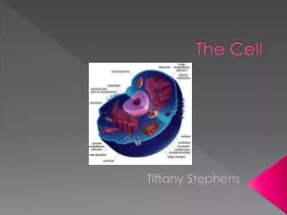
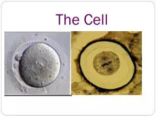
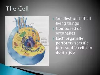
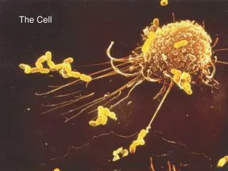
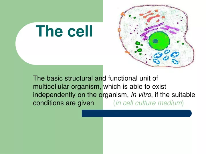
The cell The basic structural and functional unit of multicellular organism, which is able to exist independently on the organism, in vitro, if the suitable conditions are given (in cell culture medium)
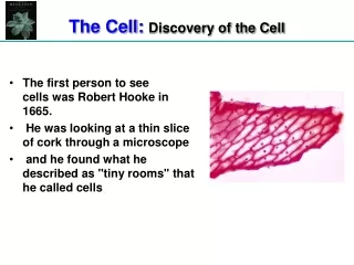
Important events in the discovery of cells • 1665 - Robert Hooke looks at cork under a microscope. Calls the chambers he see "cells" • 1665 - 75 Anton van Leeuwenhoek, the inventor of the microscope, studies organisms living in pond water. He calls them "Animalcules." • 1830 - German scientists Schleiden and Schwann summarize the findings of many scientists and conclude that all living organisms are made of cells. This forms the basis of the Cell Theory.
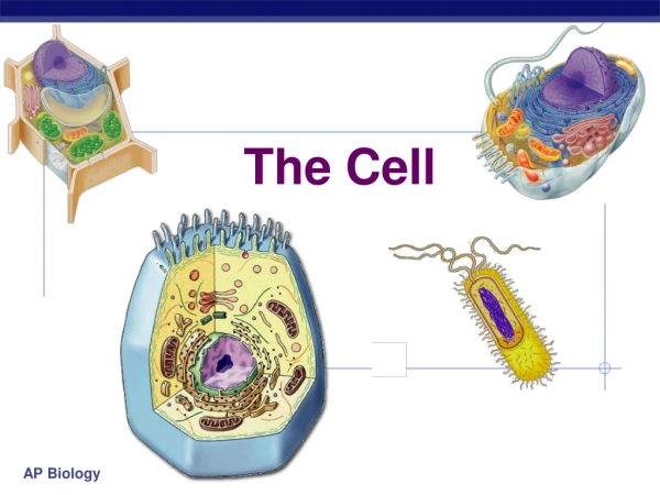
Prokaryotes Pro = before; karyon = nucleus relatively small - 5 to 10 um lack membrane-bound organelles earliest cell type Eukaryotes Eu = true; karyon = nucleus contain membrane-bound organelles Evolved from prokaryotes by endosymbiotic association of two or more prokaryotes Include Protists, Fungi, Animals, and Plants Types of Cells
The size of the cells • 5 – 120 m - granular neurons of cerebellar cortex 4 – 5 m - erythrocytes 7,4 m - Purkynje cells of cerebellar cortex or pyramidal cells of brain cortex 80 – 100 m - oocyte 120 m - megakaryocyte in bone marrow up to 150 m The majority of the somatic cells has about 10 – 20 m
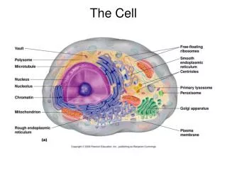
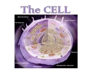
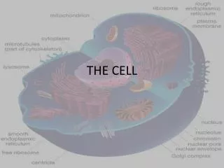
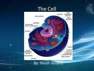
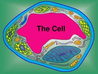
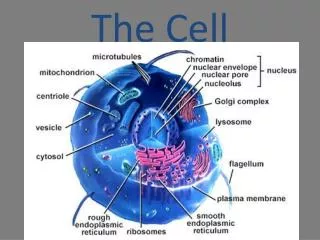
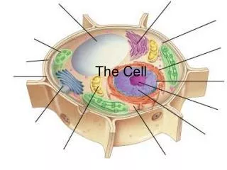
The structure of the cell Cell membrane cytosol: CELLcytoplasm organelles cytoskeleton Protoplasm inclusions nucleus nucleus is not organelle!
Cytosol = basic cytoplasm Dual colloid system: gel and sol(varies depending on the density of cytoskeleton and organelles) composition: 60 % water, 5 % minerals, 35 % organic substances (saccharids, lipids, proteins – albumins, globulins, aminoacids, phospho- lipoproteins).
The cell membrane (plasmalemma) • biomembrane – membrane units: phospholipids, proteins, cholesterol • glycocalyx • thickness 7.5 – 10 nm Cytoplasmic membrane
„fluid mosaic model“ • in EM – 2 layers of phospholipids with 3-layered appearance
Functions of integral proteins in membrane • Pumps (aktive transport, needs energy) • Canals (selectivení regulation of substances transport) • Receptors (specific bonds of molecules) • Transducers (transfer of informations into the cell) • Enzymes (on the mitochondrial membrane) • Structural proteins
Function of the cell membrane • selective barrier – regulation of substances transport from/into the cell • regulatory and recognazing functions (receptors, glycocalyx – antigenic functions)
Nucleus • controles cell activity, which is encoded in chromosomes • Numbers of nuclei in the cell (usually: 1, hepatocytes: 2, osteoklasts: 50, skeletal muscle cell: 20 - 40/1 mm of the length,human erythrocytes – without nucleus) • Size of nucleus (in many cells 5 – 15 m) • Shape of nucleus(corresponds to the cell shape – usually spherical or oval; can be lobated, segmented) • Appearance of nucleus
Nucleus structure • Nuclear envelope – consists of 2 membranes • Nuclear matrix – nucleoplasm • Chromatin (during interphase) / chromosomes (during cell division) • Nuclear skeleton • Nucleolus (1 or more)
outer nuclear membrane (+ ribosomes) perinuclear space (40 – 70 nm width) inner nuclear membrane pores (60 – 70 nm , with diaphragm and central granule) Nuclear envelope
Heterochromatin: • marginal • karyosomes Outer membrane (with ribosomes) Inner membrane Euchromatin N U C L E U S Nuclear pore Nucleolus rER (rough endo- Plasmic reticulum)
Nucleus - pores nucleus cytoplasm freez-fracture method
Nuclear matrix and skeleton • Matrix – amorphous substance surrounding chromatin and nucleolus • Composition: proteins, metabolits, ions ______________________________ • Skeleton – anastomosing trabecules
Chromatin Decondensed chromosomes during interphase • Heterochromatin – dark (spiralised and dehydrated parts of chromosomes)- marginal heterochromatin - karyosomes - perinucleolar heterochromatin (assotiated with nucleolus) • Euchromatin – pale, unstained (active parts of chromosomes with intensive synthesis of RNA)
Heterochromatin: 1.marginal, 2. karyosomes, 3. perinucleolar 2 1 3
Chromosomes • visible fibers of DNA during mitosis • Chromatids (2) • Centromere primary constriction • Organizer of nucleolus secondary constriction • Diploid set of chromosomes (2x23)in every somatic cell • Gamets – haploid set 22 + X or Y in spermatozoon 22 + X in ovum
Nucleolus • Number: not constans (1 or more), disapeare(s) during prophase of mitosis, apear(s) during telophase • Size: 1 – 2 m • Shape: round • Composition:RNA, proteins, DNA • without membrane
pars granulosa – RNA granules (preribosomes) 15 – 20 nm, pars fibrosa – RNA fibrils 3 – 5 nm, Nucleolar organizer (fibrilar center)– DNA Structure of the nucleolus
Types of nucleoli • reticular • compact • ring-shaped
Function of nucleus and nucleolus • Regulation of cell activity by RNA production ( proteosynthesis) • Communication with cell through nuclear pores • Place of genetic information (DNA), control of cell division and transfer of genetic information to daughter cells • Nucleolus – production of ribosomes (cells with intense proteosynthesis)
Memebranous Mitochondria Endoplasmic reticulum Golgi apparatus Lysosomes Peroxysomes Without membrane Ribosomes Centrioles Cell organelles
Mitochondrion • Shape: round, oval (elongated - fibrilar) • Size: 0,5 m, length of fibrilar Mi – up to 10 m • Number: different, according to metabolic activity of the cell and its energetic requirements (e.g. liver cell contains about 1000 – 2000 Mi)
Structure of mitochondrion • Outermembrane (smooth) • Innermembrane (withcristae) • Cristaemitochondriales(+ elementaryparticles) • Matrix (proteins, DNA, RNA) – semiautonomic • Mitochondrialbodies(ions) • Mitochondrialribosomes
Mitochondrial cristae Tubules Cristae Prisms
Function Mi In matrix and particles: enzymes of Krebs' cycle, oxidative phosphorylation Main function of Mi: energy releasing during ATP splitting
Ribosomes • Bodies composed of 2 subunits • Size of ribosome: 20 nm Ø free poly(ribo)somes ribosomes attached to ribosomes endoplasmic reticulum Proteosynthesis „for cell“ and „for export“ (e.g. glandular cells)
rER polysomes
Endoplasmic reticulum3D system of membranes in cell cytoplasm – 2 forms: • Granular (rough) ER – GER, RER: system of flattened, anastomosing cisterenae with (poly)ribosomes reversibly bound to membrane • Agranular (smooth) ER – AER, SER: system of tubules and vesicles with smooth membrane without ribosomes
GER – proteosynthesis (Ri) and transport of proteins into GA (by transporting vesicles) Cooperation with GA: – intracelular storing (e.g. in lysosomes and speciphic granules of leukocytes) – temporary intracelular storing before following transport from the cell (secretory granules) Function of GER
Function of AER • AER – occures in the cells which: – synthesize steroids (cells of adrenal cortex, Leydig cells of testis, cells of corpus luteum in the ovary) – break down glycogen (liver cell) – synthesize HCl (parietal cells in gastric glands) – store Ca ions (muscle cells; sarcoplasmic reticulum)
Golgi apparatus • System of smooth membranes forming 1. cisternae (5-20) 2. vesicles 3. vacuoles Polarity of GA: cis, trans cis face Transport vesicles cisternae Condensing vacuoles trans face
Transport of proteins from GER: transport vesicles Convex side – cis face (forming face) Concave – trans face (maturing face) condensing vacuoles secretory granules lysosoes Functional polarity of GA
Schema of Golgi apparatus structure „Golgi fields“
Function of GA • Postsynthetic modification of proteins (glycosylation, sulfatation, phosphorylation) • Condensation and storing of secretory products condensing vacuoles, secretory granules,lysosomes, peroxysomes • Formation of acrosomal vesicle during transformation of spermatid into spermatozoa • Donor of membranes (for some organelles)
transport vesicles