Download
1 / 52
520 likes | 530 Views
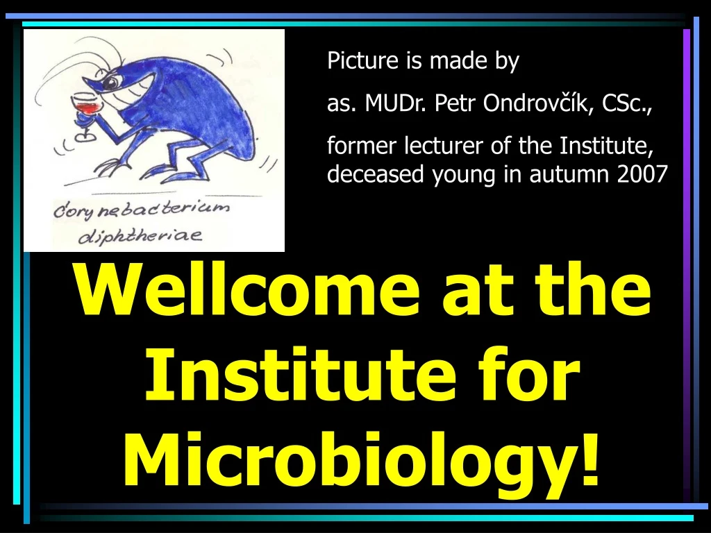
Picture is made by as. MUDr. Petr Ondrovčík, CSc., former lecturer of the Institute, deceased young in autumn 2007. Wellcome at the Institute for Microbiology!. Searching for Microbes Part I. Introduction to Diagnostics Microscopy of microbes I. Author of the slideshow: Ondřej Zahradníček

E N D
Picture is made by as. MUDr. Petr Ondrovčík, CSc., former lecturer of the Institute, deceased young in autumn 2007 Wellcome at the Institute for Microbiology!
Searching for MicrobesPart I.Introduction to DiagnosticsMicroscopy of microbes I Author of the slideshow: Ondřej Zahradníček To practicals of VLLM0421c Contacts to me: +420 777 031 969 zahradnicek@fnusa.cz ICQ 242-234-100
and tale… • Once and wolf, and tiger and HIV virus met together. • The wolf said: I wanted to bite and man, but he was armed, and so he would kill me. So I had to run away. But you, tiger, you are stronger than I am, maybe you could kill him. • The tiger said: No, no, I can‘t, the man‘s arms are stronger then I am. • Just now something laughed – something definitelly invisible. „But I‘ll get him, you‘ll see!“ It was HIV virus…
Before we shall start really • Boxes for bags and outer coats are the 1st, the 2nd and the 4th from window. Do not use the 3rd box, even when open! It is and box for secondary medical school. • Keys from boxes should be let at the table next to the computer keyboard • On the wall next to the door you can find and WC key. After coming back from WC, put it back to its place • It is forbidden to students to enter the space behind the practical‘s hall!
Safety in the laboratory – Task No. 1 • Maing risks of laboratory work are risk of fire and risk of infection • In the lab it is forbidden to eat, drink and smoke • Students are obliged to use the labcoats, but only labcoats of the Institute (NOT their own) • Students should do only what they have to do according to the protocoles and the teacher‘s bid
Literature • You need your protocoles. For this week, they are printed for you. For all remaining weeks, you will find them in Study Materials to VLLM0421c in is.muni.cz website. • You will need and textbook – Greenwood‘s one, Murray‘s one or any like these can be used. The textbook should have virological, mycological and parasitological parts – not all textbooks have this!
You can also use our websitewww.medmicro.info Photo: archive of IfM
Presence in practicals • If possible, presence in practicals should be 100 % • Rare absences (one or two) consulted before or justified afterwards are possible • Beside presence, you will alo need to forve your vigillance in practicals by and credit test or another form, that will show us that not only your body, but your soul, too, was present here
More instructions • You have to draw the pictures (draw cocci, do not write „I can see cocci“) • Draw the pictures in colours, you have to use couloured pencils • Draw what you really see (if the cocci are confluent in the microscope, draw them so • If you cannot see what you think you have to see, tell to the assistant • You will show your protocoles to Professor at the examination and they will definitelly influence your final result at the examination!
Here you can see, that fortocoles are really important (and card of and Slovak student, partially translated into English)
Classification of our branch Plant microbiology Molecular biology and genetics Infectious medicine Human medical microbiology Epidemiology of infectious diseases General microbiology Veterinary medical microbiology Dermato- venerology Cell biology
Who are clinical microbiologists Basic microbiological research Medical microbiology Other medical branches Industrial microbiology Polytechnical institutes Faculties of Science Medical faculties Your teachers are blue
A microbe (microogranism): what does it mean? • It should be living. and grain of dust is not and microbe, although it is microscopical • It should be microscopical. and giraffe is not and microbe, although it is living The second condition is not absolute. For example, and tapeworm can measure 10 m. But the eggs are microscopical, so it belongs to the microbiology.
Various microbes • Among microbes we can find microscopical algae and cyanobacteria, archea (formerly archeobacteria), various organisms able to live long time deep in the sea or in extreme conditions of hot springs • For us, clinical microbiologists, these microbes are not target of our interest. Nevertheless, they are very interesting.
What these microbes can • They live in depth of 10 km • They survive temperatures around 110 °C • They stand strong radioactivity • Instead of oxygen, they are able to „breath“ sulphur of nitrogen (or: they have another electrone acceptor than oxygen) • Nevertheless, many interesting thing are performed even by medically important microbes, as you‘ll see later.
www.meningitis.de/erreger/meningokokken.html Neisseriae
Classification of living organisms • Prions – no DNA, usually not counted to be living organisms at all • Viruses and bacteriophages • Cellullar organisms • Archaea (archeobacteria) • Eubacteria (eubacteria) • Eucarya (eukaryotic organisms) • monocellullar • polycellullar
Medically important microbes 1 • Medically important microbes are such, that are important for human body (so not for human = creator, but for human = object) • „Important for body“ is not at all the same as „harmful for body“. On the contrary, many of them are harmless, or even helping us!
Medically important microbes 2 • Each organism has its medically important microbes: human, each species of animal or plant • Even some microbes (e. g. bacteria) have their own microbes (bacteriophagi).
Neisseria gonorrhoeae http://medicine.plosjournals.org/archive/1549-1676/2/1/figure/10.1371_journal.pmed.0020024.g001-M.jpg
Main medically important microbes • Viruses (and prions) • Bacteria (e. g. and Streptococcus or an Escherichia) • Fungi (yeasts and molds) • Parasites – not all of them are microbes: • Inner parasites • Protozoa (e. g. Plasmodium malariae) • Flukes (e. g. Schistosoma haematobium) • Roundworms (e. g. Ascaris lumbricoides) • Tapeworms (e. g. Taenia saginata) • Outer parasites (lice, fleas, bugs)
Morphology of medically important microorgamisms • Viruses are composed of DNA or RNA and proeins; some viruses possess an envelope „stolen“ to a host cell • Viruses have cubic or helicoidal symetry. Several of them are able to form „pseudocrystals“ • Yeasts are egg shaped, they can form buds and so named pseudomycelia. On the surface they have a cell wall • Filamentous fungi and parasites are very variable in their shapes and they have various development stages
Morphology of bacteria • Cocci in couples (diplococci), in chains and clusters (do not say „streptococci“ and „staphylococci“, it would be confusing) • Rods straight or curved (vibria), eventually several times curved (spirillae), short or long, forming filaments or branched filaments; their ends may be round or edged and also rods may be arranged in various way • Spirochets – thin spiral bacteria • Amorph bacteria, e. g. mycoplasms (they do not have any wall, so do not have shape)
Cocci in chains (electronoptic microphotograph of Enterococcus sp.) http://www.morgenwelt.de/typo3temp/5ce14d39b5.jpg
Several times curved rods – Helicobacter http://vietsciences.free.fr/nobel/medecine/images/helicobacter%2520pylori.JPG
www.primer.ru/std/gallery_std/treponema.htm Spirochets http://nl.wikipedia.org/wiki/Afbeelding:TreponemaPallidum.jpg
Fimbriae and a flagella • Many bacteria are able to move • They move mostly by a flagella • Fimbriae are used to movement, adhesion and to excange of genetical information • Bacterial flagellae are different from flagellae of eukaryotic organisms
Bacteria with flagellae (Escherichia coli) http://www.biotox.cz/toxikon/bacteria/bacteria/obr/escherichia_coli_1.htm
http://www.laek-rlp.de/erg_pneumok.htm Capsulla and biofilm • Capsulla surrounds an individual bacterium or a couple of bacteria. It is not an integral part of bacterial cell, rather complex of molecules (mostly polysacharids), protecting the cell • Biofilm is an integral layer formed by bacteria, their capsullae and other material. It is much more resistant than an individual bacterium living in so named planctonic form
Sporulation • Sporulation is something like winter sleep, but much stronger • Spores can survive high temperatures, drying, disinfection and so on • A spore is formed as an endospore: cell is divided, but not entirelly: one part is transformed into a spore, that comes inside the second part • Bacterial spores × fungal spores!
Spores of various species of genus Bacillus http://membres.lycos.fr/neb5000/BacteriologieI/Groupes%20Bacteriens/Batonnets%20et%20coque%20Gram-positifs%20formant%20des%20endospores.htm
Spores are biochemically inactive, they do not stain in any staining method www.cropsoil.uga.edu/~parrottlab/Bugs/index.shtml.
Diagnostics: detection of bacteria and their determination • Practical medical microbiology means that a doctor (general pactitioner, specialist, doctor from hospital) sends a specimen to the lab • A laboratory of medical microbiology has to prove eventual presence of bacteria in such a specimen, and eventually to determine them. • The determination does not need to be perfect, but it has to give enough information for treatment
Specimen versus strain I: specimen Specimen is what is taken from the pacient and comes for laboratory examination • liquid or solid material in a test tube or other test tube (blood, serum, urine...) • cotton swab, usually in trasport medium.
Specimen versus strain II: strain Strain is pure culture („cultivate“) of one species of a microbe Strain can be gained only by cultivation of a microbe on a solid medium. Koch‘s discovery, that bacteria can be cultured like that, was essential for modern microbiology.
Survey of methods • Direct methods: We search for a microbe, its part or its product (e. g. a bacterial toxin) • Direct detection in specimen – we use the whole specimen (blood, urine, CSF etc.) • Strain identification – isolate determination • Indirect methods: We search for antibodies. An antibody is neither a part nor a product of a microbe – it is a macroorganism product, after being challenged by a microbe
Survey of direct methods *but in molecular epidemiology – detection of simillarity of strains - yes
Microscopy • We observe microbes, in specimen also cells of host organism(epitheliae, WBCs etc.) • Wet mount – for large and/or motile microbes (parasites, fungi, motile bacteria) • Dark field wet mount (mainly spirochets) • Fixated and stained preparations – Gram staining, Giemsa staining, Ziehl Neelsen staining (use for various groups of bacteria, fungi, parasites) • Electron microscopy – in viruses; rather for research than for common virological diagnostics
Microscopy of a specimen Microscopy of a strain Photo O. Zahradníček
Preparing a microscopical preparation • We make a smear of a swab made by a cotton swab (in stained preparations only) • Liquid specimen are dropped on a slide • If we have a strain, we make a drop of physiological saline onto the slide. We sterilize a microbiological loop in flame and after drying we take a little of bacterial mass. We mix it in a drop of saline.
Wet mount – task 2 (and + b) • In case of a wet mount a drop, in which there is a specimen or mixed strain, we do not dry. We only cover the preparation by a coverslip and we observe by objectives, magnifying e. g. 4 ×, 10 ×, 20 × or 40 ×. • We use no immersion oil • New part – task 2b: drop on slide a drop of a C..A..T. medium with pacient‘s swab. Observe microbes, but also epithelia, eventually leukocyty.
An example of a wet mount C. A. T. http://www.kcom.edu/faculty/chamberlain/Website/lectures/lecture/image/clue3.jpg
Preparing a stained preparation – task 3 • We start again by a drop of specimen or of a strain mixed in saline. In this case, the smaller the drop is, the better. • A drop is let to dry. It is allowed to help drying by placing near to the burner. • After drying, the preparation is fixated by drawing the slide throught the flame of the burner. It is necessary to check the temperature by your hand.
Simple staining • May be used if necessary directly by a general practicioner, that is far from the nearest microbiological laboratory and has methylene blue and a microscope. • Not commonly used in practice • Shows us size, shape and arrangement of microbes
Simple staining – how to do it • Fixated preparation place on a grid in sink and pour methylene blue on it • Let is be some two minutes • Then wash by tap water, and dry by filtration paper • Add a small drop of imersion oil and observe by an objective magnifying 100 ×
Simple staining Drying with filtration paper
The result may look like this(yeasts): http://biology.clc.uc.edu/fankhauser/Labs/Microbiology/Yeast_Plate_Count/09_Yeast_Meth_Blue_P7201177.jP7201179.jpg