Download
1 / 112
1.23k likes | 1.92k Views
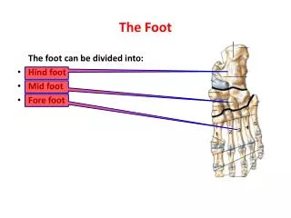
Chapter 18: The Foot. Jennifer L. Doherty, MS, LAT, ATC Academic Program Director, Entry-Level ATEP Florida International University Acute Care and Injury Prevention. Review of Anatomy. Arches of the Foot. Plantar Fascia. Joints and Ligaments of the Foot.

E N D
Chapter 18: The Foot Jennifer L. Doherty, MS, LAT, ATC Academic Program Director, Entry-Level ATEP Florida International University Acute Care and Injury Prevention
Foot Biomechanics • Foot, ankle, and leg segments form a kinetic chain • Movement of one segment effects proximal and distal segments • Entire kinetic chain must be considered when evaluating an injury • Biomechanical factors must be considered when pain occurs during walking and running
Normal Gait Two phases: • Stance or support phase • Begins at initial heel strike • Ends at toe-off • Swing or recovery phase • Represents time from toe-off to heel strike
Stance Phase • Involves weight bearing in closed kinetic chain • Five periods • Initial contact (double limb support) • Loading response (double limb support) • Mid stance (single limb support) • Terminal stance (single limb support) • Pre swing • Swing Phase • Period of non-weight bearing • Three periods • Initial swing • Mid swing • Terminal swing
Running vs. Walking • Gait patterns for running and walking have same components • During running gait… • Loading and mid-stance = more rapid • After toe off = period of no ground contact • Stance phase = 33% of running gait cycle • Accounts for 60% of walking gait cycle
At heel strike, the foot serves as shock absorber to adapt to uneven surfaces during the stance phase • Running: lateral aspect of foot makes contact with the surface placing the subtalar joint in supination • At push-off, foot serves as rigid lever to provide propulsive force • Runners: • Distance runners follow heel strike pattern • Sprinters tend to be forefoot strikers
Upon initial contact with the surface, external rotation of the tibia occurs with subtalar supination • As loading occurs, the foot and subtalar joint pronates and internal rotation of the tibia occurs • Pronation allows for unlocking of midfoot, which allows for shock absorption • Pronation also provides for even distribution of forces throughout the foot • At toe-off, the foot supinates which locks the midfoot creating a lever formation to produce greater force
Subtalar Joint Pronation and Supination • Excessive, or prolonged, pronation and supination can contribute to overuse injuries • Subtalar joint allows foot to make stable contact with ground and get into weight bearing position • Excessive motion = compensation for structural deformities, such as… • Excessive pronation: forefoot and rearfoot varus • Excessive supination: forefoot valgus
Excessive Pronation • Major cause of stress injuries • Overload of structures during stance phase • Prolonged pronation into propulsive phase • Results in loose foot • Excessive midfoot motion • Decreased stability of first ray • Increased pressure on metatarsals • Increased tibial rotation at knee
Excessive Pronation • Causes weakness push off • Does not allow foot to resupinate to provide rigid lever • Less powerful, less efficient force produced • Common injuries: • 2nd metatarsal stress fracture, Plantar fascitis Posterior tibialis tendinitis, Achilles tendinitis, Tibial stress syndrome, and Medial knee pain
Excessive Supination • Results in rigid foot • Decreased mobility of calcaneocuboid joint • Decreased mobility of first ray causing weight absorption on 1st and 5th metatarsals • Increased tension of peroneus longus • Inefficient shock absorption • Common injuries: • Inversion sprains, Tibial stress syndrome, Peroneal tendinitis, IT-Band friction syndrome, and Trochanteric bursitis
Prevention of Foot Injuries • Select appropriate footwear • Correct biomechanical structural deficiencies • Orthotics • Foot hygiene
Appropriate Footwear • Select a rigid shoe for pronators • Select a flexible shoe with additional cushioning for supinators • Other considerations: • Midsole design: controls motion along medial aspect of foot • Heel counters: controls motion in rearfoot • Outsole contour and composition • Lacing systems • Forefoot wedges
Orthotics • Utilized to correct biomechanical problems in the foot • May be constructed of… • Plastic • Rubber • Cork • Leather • Can be prefabricated or custom fitted
Foot Hygiene • Keep toenails trimmed correctly • Shave down excessive calluses • Keep feet clean • Wear clean socks and shoes that fit correclty • Keep feet as dry as possible • Prevents development of athlete’s foot
Foot Assessment History • Generic history questions • What, when, where, how??? • Questions specific to the foot • Location of pain - heel, foot, toes, arches? • Training surfaces or changes in footwear? • Changes in training, volume or type? • Does footwear increase discomfort?
Foot Assessment Observations • Does athlete favor a foot, limp, or unable to bear weight? • Does foot color change while weight bearing? • Is there pes planus/cavus? • How is foot alignment? • Are there structural deformities? • Shoe wear patterns? • Excess pronation = wear under 2nd metatarsal • Excess supination = wear on lateral border
Medial calcaneus Calcaneal dome Medial malleolus Sustentaculum tali Talar head Navicular tubercle First cuneiform First metatarsal and metatarsophalangeal joint First phalanx Lateral calcaneus Lateral malleolus Sinus tarsi Peroneal tubercle Cuboid bone Styloid process Fifth metatarsal Fifth metatarsalphalangeal joint Fifth phalanx Foot Assessment Palpations
Second, third and fourth metatarsals, metarsophalangeal joints, phalanges Third and fourth cuneiform Metatarsal heads Medial calcaneal tubercle Sesamoid bones Tibialis posterior Flexor hallucis longus Flexor digitorum longus Deltoid ligament Calcaneonavicular ligament Medial longitudinal arch Plantar fascia Transverse arch
Anterior talofibular ligament Calcaneofibular ligament Posterior talofibular ligament Peroneus longus tendon Peroneus brevis tendon Peroneus tertius Extensor hallucis longus Extensor digitorum longus tendon Extensor digitorum brevis tendon Tibialis anterior tendon
Pulses • Must ensure proper circulation to foot • Dorsalis pedis pulse • Located between extensor digitorum and hallucis longus tendons • Posterior tibial pulse • Located behind medial malleolus along Achilles tendon
Foot Assessment Special Tests • Movement • Extrinsic and intrinsic foot muscles should be assessed for pain, AROM, PROM, RROM • Tinel’s Sign • Tap over posterior tibial nerve • Positive test = tingling distal to area • Indicates presence of tarsal tunnel syndrome
Morton’s Test • Transverse pressure applied to heads of metatarsals • Positive test = pain in forefoot • Indicate presence of neuroma or metatarsalgia
Neurological Assessment Reflexes Tendon reflexes should elicit a response Achilles reflex should be assessed for the foot Sensation Cutaneous distribution of nerves must be tested Sensation can be tested by running hands over all surfaces of foot and ankle Foot Assessment
Etiology Lateral fracture MOI: severe inversion/dorsiflexion force Medial fracture MOI: severe inversion/plantarflexion force with tibial external rotation Signs and Symptoms History of repeated ankle trauma Pain with weight bearing Intermittent swelling Catching/snapping Talar dome tender upon palpation Foot Injuries Fracture of the Talus
Foot Injuries Fracture of the Talus cont. • Management • X-ray required for diagnosis • Placed on weight bearing progression • Rehab focuses on ROM and strengthening • If conservative management unsuccessful, surgery may be required • Return to play in 6-8 months following surgery
Etiology Occurs from jump or fall from height Often results in avulsion fractures anteriorly or posteriorly May present as posterior tibialis tendinitis Signs and Symptoms Immediate swelling Pain and inability to bear weight Minimal deformity unless comminuted fracture occurs Foot Injuries Fractures of the Calcaneus
Foot Injuries Fractures of the Calcaneus cont. • Management • RICE immediately • Refer for X-ray diagnosis • For non-displaced fracture, immobilization and early ROM exercises when pain and swelling subside
Etiology Occurs due to repetitive trauma Characterized by sudden onset of pain in plantar-calcaneal area Signs and Symptoms Weight bearing (particularly at heel strike) causes pain Pain continues following exercise May require bone scan for diagnosis Foot Injuries Calcaneal Stress Fracture
Foot Injuries Calcaneal Stress Fracture cont. • Management • Conservative for 2-3 weeks • Including rest and AROM • Non-weight bearing cardio training should continue • As pain subsides, activity can be returned gradually
Etiology Traction injury at apophysis of calcaneus Where Achilles tendon attaches to calcaneous Signs and Symptoms Pain occurs at posterior heel below Achilles attachment Pain occurs during vigorous activity Pain ceases following activity Foot Injuries Apophysitis of the Calcaneus (Sever’s Disease)
Foot Injuries Apophysitis of the Calcaneus (Sever’s Disease) cont. • Management • Best treated with ice, rest, stretching and NSAID’s • Heel lift could also relieve some stress
Etiology Caused by inflammation of bursa beneath Achilles tendon Result of pressure and rubbing of shoe heel counter Chronic condition that develops over time May take extensive time to resolve Exostosis may also develop Signs and Symptoms Pain with palpation superior and anterior to Achilles insertion Swelling on both sides of the heel cord Foot Injuries Retrocalcaneal Bursitis (Pump Bump)
Foot Injuries Retrocalcaneal Bursitis (Pump Bump) cont. • Management • RICE and NSAID’s used as needed • Ultrasound can reduce inflammation • Routine stretching of Achilles • Heel lifts to reduce stress • Donut pad to reduce pressure • Possibly invest in larger shoes with wider heel contours
Etiology Caused by sudden starts, stops or changes of direction Irritation of fat pad Pain often on the lateral aspect due to heel strike pattern Sign and Symptoms Severe pain in heel Unable to withstand stress of weight bearing Often warmth and redness over the tender area Foot Injuries Heel Contusion
Foot Injuries Heel Contusion cont. • Management • Reduce weight bearing for 24 hours • RICE and NSAID’s • Resume activity with heel cup or doughnut pad after pain has subsided • Wear shock absorbent shoes
Etiology Pronation and trauma resulting in displacement of the cuboid Often confused with plantar fascitis Pain due to stress on long peroneal muscle with the foot in pronation Signs and Symptoms Pain along 4th and 5th metatarsals Pain over the cuboid May refer pain to heel area Pain may increase following long periods of weight bearing Foot Injuries Cuboid Subluxation
Foot Injuries Cuboid Subluxation cont. • Management • Dramatic results may be obtained with joint mobilization • Orthotic can be used to maintain position of cuboid
Foot Injuries Tarsal Tunnel Syndrome • Tunnel behind medial malleolus • Osseous floor • Roof composed of flexor retinaculum • Etiology • Any condition that compromises tibialis posterior, flexor hallucis longus, flexor digitorum, and tibial nerve, artery, or vein • May result from previous fracture, tenosynovitis, acute trauma, or excessive pronation
Foot Injuries Tarsal Tunnel Syndrome cont. • Signs and Symptoms • Pain and paresthesia along medial and plantar aspect of foot • Motor weakness and atrophy may result • Increased pain at night • Positive Tinel’s Sign • Management • NSAID’s and anti-inflammatory modalities • Orthotics • Possibly surgery if condition is recurrent
Etiology Foot is hyperplantarflexed when foot is already plantaflexed Rearfoot is locked resulting in dorsal displacement of metatarsal bases Signs and Symptoms Pain Inability to bear weight Swelling Tenderness localized on dorsum of foot Possible metatarsal fractures Sprains of 4th and 5th tarsometatarsal joints May cause severe disruption of ligaments Foot Injuries Tarsometatarsal Fracture Dislocation (Lisfranc Injury)