Download
1 / 28
310 likes | 533 Views
Week 2 (3/12-3/16) Sub-Cloning. Week 1 (this week) Fluorescence labeling and microscopy: The actin and tubulin cytoskeleton in cultured cells. Week 3 (3/19-3/23) Transfection & vital staining: The secretory and endocytic pathways. Overview of 130L Part 2.

E N D
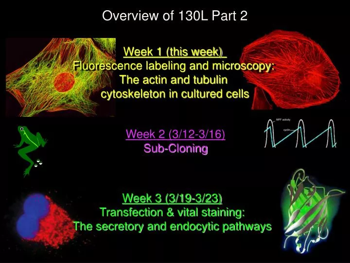
Week 2 (3/12-3/16) Sub-Cloning Week 1 (this week) Fluorescence labeling and microscopy: The actin and tubulin cytoskeleton in cultured cells Week 3 (3/19-3/23) Transfection & vital staining: The secretory and endocytic pathways Overview of 130L Part 2
What you should get from today’s lecture • What are cultured cells and why do we use them? • General background on the cytoskeletonand the drug treatments used in this lab • How are cells fixed and stained with fluorescent reagents? • An introduction to microscope resolution
single cell (scanning EM) colony of cells dish of cell colonies 100 mm >0.01 mm 1 mm How big are animal cells?
Cells in culture can undergo diverse differentiation pathways Neuronal cell extending axons and dendrites Mouse C2C12 muscle cells forming myofibers
embryonic stem cells “feeder” cells Stem cells are undifferentiated and pluripotent, meaning that they can differentiate to become multiple other cell types
Cell culture= propagation of cells outside the organism The Good: • Cellular environment can be easily observed and manipulated • a. Injection • b. Transfection -introduction of genes • c. Pharmaceutical or genetic manipulation (RNAi) • d. Fluorescent tracers (live and fixed cells) • 2) Cell type can be well defined (How?) • 3) Large quantities of cells can be obtained - biochemistry • 4) Diverse cellular functions can be investigated • 5) Noninvasive way to study mammalian cells The Bad: 1) Requires care and $$$ to grow & prevent contamination 2) May not always recapitulate real cellular physiology
Most cells in the body or removed from the body are MORTAL:they have a finite ability to replicate and divide Immortal cell lines can arise by spontaneous mutation or deliberate transformation of mortal cell strains. Embryonic stem cells are also immortal. We don’t really understand why they have this unlimited replication potential, or why most cells lack it.
Lab: Visualizing the cytoskeleton using fluorescence microscopy Purpose: stain cells to observe the cytoskeleton; Observe and record the effects of different drugs on cytoskeletal components and cell shape. 2 cytoskeletal components will be examined: Actin - required for cell shape & movementsincluding translocation and cytokinesis Tubulin - forms microtubule “tracks” that enable chromosomes, vesicles, etc. to move in targeted ways within cells. Your cells will be treated with 4 drugs (+ controls) TPA/PMA Latrunculin* Taxol (Paclitaxel)* Nocodazole* *alter the equilibrium between subunits and polymersof actin or tubulin
Cells move! all cell movement requires actin dynamics
actin filaments form three-dimensional branching networks ARP 2/3 complex actin monomer
mimics 1,2 diacylglycerol (DAG) • DAG plus Ca++ activate protein kinase C (PKC) • PKC activation results in phosphorylation of MANY PKC targets (i.e. MARCKS myristolated alanine rich C kinase substrate) • this leads to major changes in cell growth, cell shape and the cytoskeleton TPA = phorbal myristate acetate = PMA Phorbol ester
Latrunculin A free G-actin G-actin bound to Latrunculin A
Microtubules DNA Microtubules control other aspects of cell dynamics including vesicle transport and chromosome segregation interphase mitosis
Microtubule dynamics are controlled by tubulin conformation, which can be modified by other proteins or drugs such as nocodazole or taxol Eva Nogales’ lab
Microtubule dynamics at the cell edge Individual microtubules are constantly growing and shrinking. Their rate of growth and the frequency of switching between growth and shrinkage are controlled by a large number of factors, including MAPs (microtubule associated proteins).
Taxol binds to polymerized β-tubulin and inhibits depolymerization Tubulin α/β dimer taxol GTP/GDP
Cell Fixation and Permeabilization A. Chemical fixation - kills and immobilizes cells • Aldehydes (formaldehyde, glutaraldehyde): Cross-link amino groups in proteins. • Stabilizing many structures • Can block antibody access to targets. • Alcohols (methanol or ethanol, with or without acetic acid):Denatures and precipitates proteins in place. Alcohol fixation does not retain soluble proteins • Cell/protein morphology not preserved Fast and easy • Good for some labile structures (microtubules)
Cell Fixation and Permeabilization • Permeabilization - detergents are used to solubilize cellmembranes to allow staining reagents to penetrate;makes proteins accessible to staining reagents. • Usually gentle, non-ionic detergents are used: Brij-58 Tween-20 Triton X-100 • Effect of detergents can depend upon order of steps (as in this week’s lab): • When added before fixative, they will often solubilize proteins (e.g. unpolymerized tubulin, as in today’s lab). • This can be a technical advantage - reduces background - but can also lead to experimental artifacts.
Staining of Actin Phalloidin:a natural toxin from some mushrooms Binds to filamentous actin (F-actin) only For this lab, we buy phalloidin that is covalently linked to rhodamine Phalloidin + Amanita phalloides TRITC = tetramethyl rhodamine isothiocyanate Kidney cellstained withrhodamine-phalloidin
Variable regions Antibodies are highly conserved molecules with variable regionsthat specify antigen recognition and affinity Constant region Antibody, or Immunoglobulin
Fluorescein, or FITC Secondary antibodies(react with primary antibodies) Antibodies are highly conserved molecules with variable regionsthat specify antigen recognition and affinity
Fluorescein Tubulin immobilizedby cell fixation Secondary antibodies:Goat antibodies raised against mouse antibodies (IgGs),conjugated to a fluorophore (fluorescein) Primary antibodies:mouse antibodies raised against antigen (tubulin) Staining of Microtubules using“Indirect Immunofluorescence” 1o Ab: mouse anti-tubulin; 2o Ab: fluorescein goat anti-mouse Note: color of the fluorescence isdetermined by the fluorophore attachedto the secondary antibody!
DAPI and Hoechst are dyes that bind directly to DNAand fluoresce brightly, with very similar spectra DAPI (diamidino phenyl indole) Hoechst33258/33342 DAPI bound to the minor groove