Download
1 / 59
620 likes | 1.01k Views
Chapter 19. The Blood. The Blood. The cardiovascular system consists of three components: Blood Heart Blood vessels. Functions of Blood. Blood transports: oxygen & CO2 between lungs & tissues nutrients from the digestive tract metabolic wastes- from body to kidneys for elimination

E N D
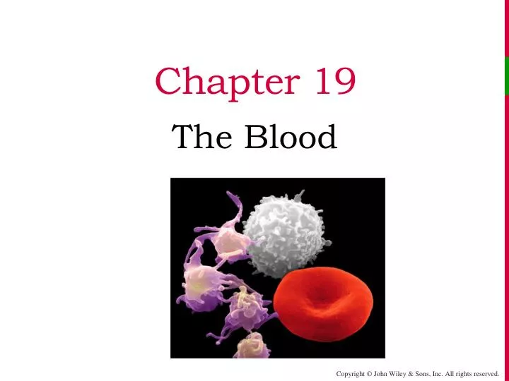
Chapter 19 The Blood
The Blood • The cardiovascular system consists of three components: • Blood • Heart • Blood vessels
Functions of Blood • Blood transports: • oxygen & CO2 between lungs & tissues • nutrients from the digestive tract • metabolic wastes- from body to kidneys for elimination • hormones from endocrine glands to target organs • Blood regulates: • body temperature • pH using buffer systems • Blood protects • against blood loss by initiating hemostasis & coagulation • against infection – antibodies, complement proteins, WBCs
Physical Characteristics of Blood • It is viscous (thick) and more dense than water • temperature slightly warmer than core body temperature (T = 38o C) • slightly alkaline pH (7.35-7.45) • oxygenated blood bright red, poorly oxygenated blood dark red • makes up 8% of body mass • Blood volume: 5–6 L for males, and 4–5 L for females
Constituents of Blood • Blood is 5 liters of a specialized fluidconnective tissue composed of formed elements (45%) suspended in a solution called plasma (55%) • If a sample of blood is centrifuged cellular portion will precipitate out of solution and form a heavier sediment below the straw colored liquid plasma • The normal RBC mass is almost 45% by volume – this is called the hematocrit (Hct)
Blood Plasma • Plasma is 92% water, with dissolved solutes: • Plasma proteins – produced by the liver • albumin, globulins, fibrinogen • Nitrogenous wastes • urea, uric acid, creatinine • Nutrients – • glucose, fatty acids, amino acids • Electrolytes • sodium, potassium, calcium, chloride, bicarbonate • Respiratory gases • oxygen and carbon dioxide • Hormones • Clotting factors • If plasma is allowed to coagulate it is called serum - serum is just plasma without the clotting factors- used or blood testing
Plasma Proteins • Albumin • contributes significantly to colloidal osmotic pressure of blood • It also plays an important role as a carrier molecule for lipid soluble substances e.g. hormones • globulins, of which there are several types: • α-globulins and β- globulins are carrier proteins • δ-globulins are immunoglobulins (antibodies) made by activated B lymphocytes called plasma cells • Fibrinogen- clotting factor –forms the blood clot
Formed Elements • The formed elements of blood include: • Erythrocytes ( RBCs)- highest count • Leukocytes( WBCs) • Platelets • Are continuously formed in the bone marrow
Hematopoiesis • The process by which the formed elements of blood develop is called hemopoiesis (hematopoiesis). • blood cells are formed in red bone marrow from pluripotent stem cells. • Red bone marrow: • In adults : bones of axial skeleton, pectoral & pelvic girdles, heads of humerus & femur • In newborns- all bone marrow is red
Hematopoiesis • Blood cells are formed from pluripotent stem cells. • The pluripotent stem cells (hemopoietic stem cells) produce the myeloid stem cell and lymphoid stem cell • myeloid stem cell gives rise to: RBCs, platelets, monocytes, neutrophils, eosinophils, and basophils • lymphoid stem cell gives rise to: T lymphocytes, B lymphocytes, NK cells • Regulation of hematopoiesis: • Erythropoietin (EPO) regulates RBC formation • Thrombopoietinregulates platelet formation • Colony stimulating factors & interleukins stimulate WBC formation
Clinical connection • Bone marrow examination by bone marrow aspiration or bone marrow biopsy • For diagnosis of leukemias, anemias • Site: usually iliac crest of hip bone, sternum
Red Blood Cells • About 5 million RBCs/mm3 of blood • Red blood cells are bi-concave discs- • increases the cell surface area- gives them a high oxygen carrying capacity • Mature RBCs don't have a nucleus or any protein making machinery -die in about 120 days. • specific purpose – to carry O2 to the tissues of the body- also carry 23 % CO2 • Their shape also allows them to deform and fit in small capillary beds
RBCs and Hemoglobin • Each RBC contains 280 million molecules of Hb • Hemoglobin (Hb) is a protein molecule adapted to carry O2 (and CO2 as well) • A Hgb molecule consists of globin protein (2 alpha and 2 beta polypeptide chains), each embedding an iron-containing heme group • Oxygen binds to the heme group ( one Hb binds 4 oxygen molecules) • CO2 binds to globin proteins
RBCs • RBCs and CO2: • Red blood cells contain the enzyme carbonic anhydrase • Carbon dioxide diffuses fron tissues into RBCs • It combines with water to form carbonic acid- H2CO3 which quickly dissociates into hydrogen ions and bicarbonate ions • 70 % of CO2 is transported in blood as bicarbonate ions which is an important blood buffer
RBCs Life Cycle • Erythropoiesis is the part of hematopoiesis that deals with the production of RBCs. • Control is by negative feedback • Erythropoiesis increases when states of hypoxia (O2 deficiency)- stimulates the kidneys to release the hormone erythropoietin (EPO) • EPO circulates to the red marrow and speeds up the maturation and release of immature red cells
Negative feedback regulation of Erythropoiesis Some stimulus disrupts homeostasis by Decreasing Oxygen delivery to kid-neys (and other tissues) Receptors Kidney cells detect low oxygen level Input Increased erythropoietin secreted into blood Control center Return to homeostasis when oxygen delivery to kidneys increases to normal Proerythroblasts in red bone marrow mature more quickly into reticulocytes Output Increased erythropoietin secreted into blood Effectors Larger number of RBCs in circulation Increased oxygen delivery to tissues
RBCs Life Cycle • As cells mature in the bone marrow, they become smaller • the nucleus is lost • Most organelles lost • the amount of Hb increases At reticulocyte stage RBCs are sent out into the blood stream
Reticulocytes Reticulocytes: At this stage the RBCs are sent out into the blood stream Normally 1-2% of the RBCs in the peripheral circulation are reticulocytes • The rate of erythropoiesis is measured by the number of immature RBCs (called reticulocytes or “retics”) in the peripheral circulation • A low retic count (<.5%) indicates a low rate of erythropoiesis while an elevated rate (>2%) indicates a high rate of erythropoiesis
RBC Life Cycle • RBCs live only about 120 days- are destroyed mainly in spleen • To maintain normal numbers, new cells must enter the circulation at 2 million/s to balance high rate of RBC destruction • Ruptured RBCs are destroyed by macrophages in the spleen and liver • Heme and globin are separated • Globin is metabolized into amino acids- released into the circulation • Iron of the heme is salvaged for re-use • Heme is degraded to bilirubin
RBC Life Cycle Circulation for about 120 days Circulation for about 120 days Circulation for about 120 days Circulation for about 120 days Circulation for about 120 days Circulation for about 120 days 7 7 7 7 7 7 7 3 3 3 3 3 3 3 3 3 3 3 Reused for protein synthesis Reused for protein synthesis Reused for protein synthesis Reused for protein synthesis Reused for protein synthesis Reused for protein synthesis Reused for protein synthesis Reused for protein synthesis Reused for protein synthesis Reused for protein synthesis Reused for protein synthesis Amino acids Amino acids Amino acids Amino acids Amino acids Amino acids Amino acids Amino acids Amino acids Amino acids Amino acids Transferrin Transferrin Transferrin Transferrin Transferrin Transferrin Transferrin Transferrin Fe3+ Fe3+ Fe3+ Fe3+ Fe3+ Fe3+ Fe3+ Fe3+ Globin Globin Globin Globin Globin Globin Globin Globin Globin Globin Globin Globin 6 6 6 6 6 6 6 6 4 4 4 4 4 4 4 4 4 4 5 5 5 5 5 5 5 5 5 Fe3+ Fe3+ Fe3+ Fe3+ Fe3+ Fe3+ Fe3+ Fe3+ Fe3+ Fe3+ 2 2 2 2 2 2 2 2 2 2 2 2 Fe3+ Fe3+ Fe3+ Fe3+ Fe3+ Fe3+ Fe3+ Ferritin Ferritin Ferritin Ferritin Ferritin Ferritin Ferritin Ferritin Ferritin Heme Heme Heme Heme Heme Heme Heme Heme Heme Heme Heme Heme Transferrin Transferrin Transferrin Transferrin Transferrin Transferrin Transferrin Transferrin Transferrin Transferrin + + + + + + + Globin Globin Globin Globin Globin Globin Globin Bilirubin Bilirubin Bilirubin Bilirubin 9 9 9 9 9 + + + + + + + Biliverdin Biliverdin Biliverdin Biliverdin Biliverdin Liver Liver Liver Liver Liver Liver Liver Liver Liver Vitamin B12 Vitamin B12 Vitamin B12 Vitamin B12 Vitamin B12 Vitamin B12 Vitamin B12 Bilirubin Bilirubin Bilirubin Bilirubin Bilirubin 11 11 11 1 1 1 1 1 1 1 1 1 1 1 1 1 Red blood cell death and phagocytosis Red blood cell death and phagocytosis Red blood cell death and phagocytosis Red blood cell death and phagocytosis Red blood cell death and phagocytosis Red blood cell death and phagocytosis Red blood cell death and phagocytosis Red blood cell death and phagocytosis Red blood cell death and phagocytosis Red blood cell death and phagocytosis Red blood cell death and phagocytosis Red blood cell death and phagocytosis Red blood cell death and phagocytosis + + + + + + + 10 10 10 10 Erythopoietin Erythopoietin Erythopoietin Erythopoietin Erythopoietin Erythopoietin Erythopoietin Small intestine Small intestine Small intestine Kidney Kidney 8 8 8 8 8 8 Erythropoiesis in red bone marrow Erythropoiesis in red bone marrow Erythropoiesis in red bone marrow Erythropoiesis in red bone marrow Erythropoiesis in red bone marrow Erythropoiesis in red bone marrow Bilirubin Bilirubin Bilirubin 13 13 12 12 12 Urobilin Urobilin Urobilinogen Urobilinogen Urobilinogen Macrophage in spleen, liver, or red bone marrow Macrophage in spleen, liver, or red bone marrow Macrophage in spleen, liver, or red bone marrow Macrophage in spleen, liver, or red bone marrow Macrophage in spleen, liver, or red bone marrow Macrophage in spleen, liver, or red bone marrow Macrophage in spleen, liver, or red bone marrow Macrophage in spleen, liver, or red bone marrow Macrophage in spleen, liver, or red bone marrow Macrophage in spleen, liver, or red bone marrow Macrophage in spleen, liver, or red bone marrow Macrophage in spleen, liver, or red bone marrow Macrophage in spleen, liver, or red bone marrow Key: Key: Key: Key: Key: Key: Key: Key: Key: Key: Key: Key: Key: Bacteria Bacteria Bacteria in blood in blood in blood in blood in blood in blood in blood in blood in blood in blood in blood in blood in blood Stercobilin Stercobilin Stercobilin 14 Large intestine in bile in bile in bile in bile in bile in bile in bile in bile in bile in bile in bile in bile in bile Feces Feces Feces Urine Urine
Clinical connections • Anemia is a condition of insufficient RBC’s or hemoglobin (quality or quantity) • It is most often the result of low iron intake, hemolysis, blood loss, or lack of production in the bone marrow • Polycythemiais a condition of excess number of RBCs • It occurs in response to hypoxia, shots of EPO (illegal “doping”), COPD
Anemias • Iron deficiency anemia is the most common anemia in the U.S., and affects primarily menstruating women • Chronic blood loss is a cause • Hemorrhagic anemia is the result of precipitous blood loss, and results in an equal decrease in Hct, Hb content, and RBC count
Anemias • Sickle-cell disease (SCD), also called sickle-cell anemia, is an autosomal recessive disorder. A genetic defect in the primary DNA sequence leads to production of a faulty Hb β chain, and RBCs that take on a rigid, sickle-shape • Sickling decreases the cells' flexibility and results in a variety of complications; life expectancy is shortened
White Blood Cells • WBCs number- between 5000-10,000 cells/mm3 • There are 5 different types of WBCs • (WBCs)or leukocyteshave nuclei and other organelles
Leukocytes • Leukocytes are divided into two groups depending on whether they contain conspicuous cytoplasmic granules (when stained) • Granulocytes include the neutrophils, eosinophils, and basophils • Agranulocytes are the monocytes and lymphocytes
Neutrophils • The most numerous WBC in normal blood (60-70% )is the neutrophil, or polymorphonucleocyte (PMN) • PMNs are granulocytes lilac granules and nuclei have 2-5 lobes • They are phagocytes - their principal role is to fight bacterial infections PMN phagocytizing a microbe
Eosinophils • Eosinophils are characterized by their large red granules and bilobed nuclei • They are (2-4% of circulating WBCs), but their numbers increase slightly with allergic reactions & parasitic infections • they have also been associated with the development of allergies
Basophils • Basophils are the granulocytes that contain large, dark blue, histamine containing granules • Normally, they are the lowest number of circulating WBCs (only 0-1%), • May have a role to play in the inflammatory responses
Monocytes • While monocytesare not granulocytes, they come from the same immediate precursor cell as the 3 granulocytes (the myeloid stem cell) • 3-8% of the circulating WBCs • Along with neutrophils, monocytes are the other major group of phagocytic cells. • they are more numerous in the peripheral, tissues where they act as “fixed” phagocytes
Lymphocytes • Approximately 20-30% of circulating WBCs are lymphocytes • increase in number in acute viral infections • Most lymphocytes continually move between lymphoid tissues, lymph, and blood, spending only a few hours in blood Lymphocytes are the cornerstone of the specific immune response • There are two types of lymphocytes: T cells and B cells • T cells control immune responses • B cells give rise to plasma cells, which produce antibodies
Functions of Leukocytes • To get rid of pathogens the WBCs have to leave the blood stream and collect at sites of infection or injury • WBCs leave the bllod vessels by the process called emigration : • Rolling along the endothelium & then sticking- by complementary adhesion molecules on endothelium (selectins) and on leucocytes (integrins ) • Squeeze out between endothelial cells -diapedesis • Chemicals released by microbes and inflamed tissues attract phagocytes, a phenomenon called chemotaxis
Functions of Leukocytes • Neutrophils are the first to emigrate in bacterial infections • Reach by chemotaxis & engulf pathogen by phagocytosis • Release strong oxidants & defensins to destroy engulfed bacteria • Neutrophils are followed by monocytes- which get transformed into phagocytic tissue macrophages • Lymphocytes take part in immune responses • B cells- antobody mediated humoral immunity • T cells – immune regulation & destruction of viral infected and cancer cells
Interstitial fluid Blood flow Emigration of neutrophils Neutrophil Endothelial cell Rolling Sticking Squeezing between endothelial cells Key: Selectins on endothelial cells Integrins on neutrophil
Diapedesis Chemotaxis & phagocytosis From Wikimedia Commons
WBC Indices • For diagnostic purposes, physicians measure the total leucocyte count • A leukocytosisis any WBC count > 10,000/mm3, and usually indicate an infectious process or inflammation • A leukopenia is any WBC count < 5,000/mm3, and usually indicates a severe disease (AIDS, bone marrow failure, severe malnutrition, or chemotherapy) • Differential leucocyte count : percentages of each of the 5 types of WBCs
WBC Indices • Shifts in the normal percentages of circulating WBCs will often point towards a bacterial infection (elevated neutrophils) or a viral infection (elevated lymphocytes) • In this peripheral blood smear a patient with lymphocytic leukemia has a WBC >150,000 and 90% of the WBCs are cancerous lymphocytes! Lymphocytic leukemia.
Platelets • Platelets (thrombocytes) are about 150,000-400,000 cells/mm3 , they have a short life span (5 to 9 days) • Their granules contain chemicals (serotonin, Ca2+, ADP) that, once released, promote blood clotting Also release PDGF- promotes growth & healing of damaged vessels
Platelets • Megakaryocytes (immediate precursors of platelets) are huge cells that splinter into 2000 to 3000 fragments while still in the red bone marrow • Each disc shaped fragment is a platelet • Platelets leave the red bone marrow and enter the circulation
Hemostasis • Hemostasis is a sequence of responses that stops bleeding when blood vessels are damaged or ruptured • Three mechanisms reduce blood loss • Vascular spasm • Formation of a platelet plug • Blood clotting (coagulation)
Hemostasis • Vascular spasm occurs as damaged blood vessels constrict- (by neural & chemical stimuli) • Platelets adhere to damaged endothelium to form a platelet plug
Red blood cell Red blood cell Red blood cell Platelet Platelet Platelet Collagen fibers and damaged endothelium Collagen fibers and damaged endothelium Collagen fibers and damaged endothelium 1 1 1 Platelet adhesion Platelet adhesion Platelet adhesion 1 1 1 Liberated ADP, serotonin, and thromboxane A2 Liberated ADP, serotonin, and thromboxane A2 2 2 2 2 Platelet release reaction Platelet release reaction Platelet plug 3 Platelet aggregation 3 2.Platelet Plug Formation
Hemostasis • Clotting (coagulation) is possible because of the presence of several clotting proteins normally dissolved in the blood in inactive state Coagulation occurs in a cascade whereby one activated clotting protein triggers the next step in the process, which triggers the next • There are 2 pathways to activate the clotting cascade: extrinsic & intrinsic
(a) Extrinsic pathway (a) Extrinsic pathway (a) Extrinsic pathway (b) Intrinsic pathway (b) Intrinsic pathway (b) Intrinsic pathway Tissue trauma Tissue trauma Tissue trauma Blood trauma Blood trauma Blood trauma Damaged endothelial cells expose collagen fibers Damaged endothelial cells expose collagen fibers Damaged endothelial cells expose collagen fibers Tissue factor (TF) Tissue factor (TF) Tissue factor (TF) Damaged platelets Damaged platelets Damaged platelets Activated XII Activated XII Activated XII Activated platelets Activated platelets Activated platelets Ca2+ Ca2+ Ca2+ Ca2+ Ca2+ Ca2+ + + Platelet phospholipids Platelet phospholipids Platelet phospholipids Activated X Activated X Activated X Activated X Activated X Activated X V V V V V V + + Ca2+ Ca2+ Ca2+ Ca2+ Ca2+ Ca2+ 1 1 1 PROTHROMBINASE PROTHROMBINASE PROTHROMBINASE (c) Common pathway (c) Common pathway Ca2+ Ca2+ Prothrombin (II) Prothrombin (II) 2 2 THROMBIN THROMBIN Ca2+ XIII Fibrinogen (I) Activated XIII STRENGTHENED Loose fibrin threads 3 FIBRIN THREADS Blood clotting cascade
Hemostasis • The extrinsic pathway has few steps, occurs within seconds, once the protein “tissue factor” (TF) or tissue thromboplastin leaks into the blood from cells • The intrinsic pathway is more complex, & slower in response blood contact with collagen under endothelial cells- activates factor XII
Hemostasis • Both the extrinsic and intrinsic clotting pathways converge at a common point (pathway) where factor X becomes activated (Xa), combines with factor V to form: • Prothrombin activator(prothombokinase) • Prothrombin is converted into thrombin • Thrombin converts soluble fibrinogen into insoluble fibrin threads,
Hemostasis • Ca2+plays an important role throughout the clotting system • Clot retraction is the consolidation of the fibrin clot. • Fibrin threads contract as platelets pull on them • As the clot retracts, it pulls the edges of the damaged vessel closer together, decreasing the risk of further damage – new endothelial cells can then repair the vessel lining
Fibrinolysis • Because blood clotting involves positive feedback cycles, a clot has a tendency to enlarge, this is checked by: • The fibrinolytic system - dissolves clots • Vessel endothelial cells release tissue plasminogenactivator( tPA) that can activate plasminogenan inactive plasma enzyme to become plasmin, (the enzyme that actively dissolves clots)
Intravascular Clotting • Inappropriate clotting in an unbroken blood vessel is called thrombosis; the clot itself, called a thrombus • Such clots may be initiated by: • endothelial injury resulting from atherosclerosis, trauma, or infection • Stasis of blood- allowing clotting factors to accumulate locally and initiate the coagulation cascade