Download
1 / 68
680 likes | 689 Views
NINE ABDOMINO-PELVIC REGIONS. Maintaining Homeostasis. The body communicates through nervous and endocrine systems consisting of 3 basic components 1) Receptor Detects a stimulus 2) Control center Analyzes information Determines appropriate response

E N D
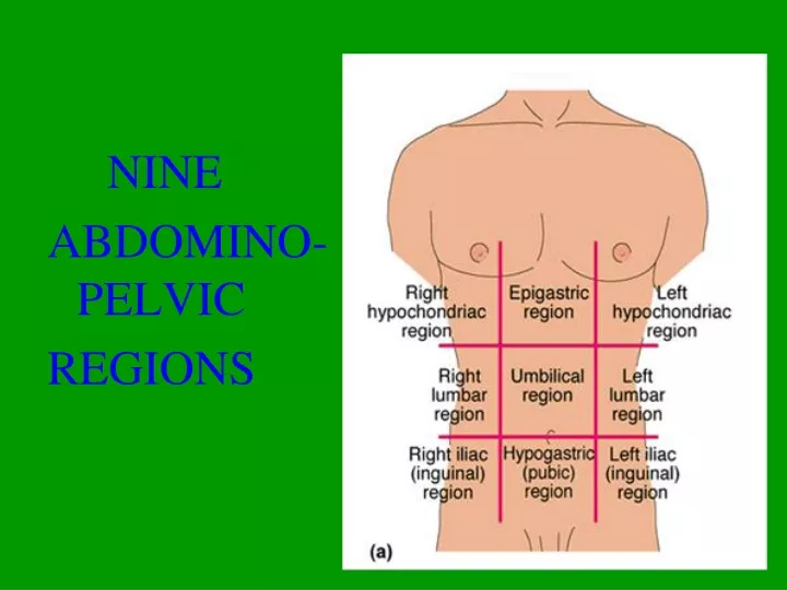
NINE ABDOMINO-PELVIC REGIONS
Maintaining Homeostasis • The body communicates through nervous and endocrine systems consisting of 3 basic components 1) Receptor • Detects a stimulus 2) Control center • Analyzes information • Determines appropriate response 3) Effector (Muscles or glands) • Responds to the stimulus
Cranium Skull Facial bones Clavicle Thoracic cage (ribs and sternum) Scapula Sternum Rib Humerus Vertebra Vertebral column Radius Ulna Sacrum Carpals Phalanges Metacarpals Femur Patella Tibia Fibula Tarsals Metatarsals (a) Anterior view Phalanges Figure 7.1a
C1 Cervical curvature (concave) 7 vertebrae, C1–C7 Spinous process Transverse processes Thoracic curvature (convex) 12 vertebrae, T1–T12 Intervertebral discs Intervertebral foramen Lumbar curvature (concave) 5 vertebrae, L1–L5 Sacral curvature (convex) 5 fused vertebrae sacrum Coccyx 4 fused vertebrae Anterior view Right lateral view Figure 7.16
Phalanges Distal Middle Proximal 1 2 3 4 5 Metatarsals Medial cuneiform Intermediate cuneiform Lateral cuneiform Navicular Cuboid Tarsals Talus Trochlea of talus Calcaneus (a) Superior view Figure 7.33a
Phalanges • Distal • Middle • Proximal Metacarpals • Head • Shaft • Base Sesamoid bones Carpals Carpals Carpals • Trapezium • Hamate • Trapezium • Trapezoid • Capitate • Trapezoid • Scaphoid • Pisiform • Scaphoid • Triquetrum Radius • Lunate Ulna Radius (a) Anterior view of left hand (b) Posterior view of left hand Figure 7.28a-b
Gouty Arthritis • Deposition of uric acid crystals in joints and soft tissues, followed by inflammation • More common in men; Typically affects the joint at the base of the great toe • In untreated gouty arthritis, the bone ends fuse and immobilize the joint • Treatment: drugs, plenty of water, avoidance of alcohol
Rheumatoid Arthritis (RA) Chronic, inflammatory, autoimmune disease of unknown cause • Usually arises between age 40 and 50, but may occur at any age; affects 3 times as many women as men • Signs and symptoms include joint pain and swelling (usually bilateral), anemia, osteoporosis, muscle weakness, and cardiovascular problems; RA begins with synovitis of the affected joint • Inflammatory blood cells migrate to the joint, release inflammatory chemicals • Inflamed synovial membrane thickens into a pannus • Pannus erodes cartilage, scar tissue forms, articulating bone ends connect (ankylosis) • Conservative therapy: aspirin, long-term use of antibiotics, and physical therapy • Progressive treatment: anti-inflammatory drugs or immunosuppressants
Movements at Synovial Joints • Gliding • Angular movements: • Flexion, extension, hyperextension • Abduction, adduction • Circumduction • Rotation • Medial and lateral rotation
Movements at Synovial Joints 4. Special movements • Supination, pronation • Dorsiflexion, plantar flexion of the foot • Inversion, eversion • Protraction, retraction • Elevation, depression • Opposition
Gliding Movements • One flat bone surface glides or slips over another similar surface • Examples: • Intercarpal joints • Intertarsal joints • Between articular processes of vertebrae
Gliding (a) Gliding movements at the wrist Figure 8.5a
Angular Movements Movements that occur along the sagittal plane: • Flexion—decreases the angle of the joint • Extension— increases the angle of the joint • Hyperextension—excessive extension beyond normal range of motion
Hyperextension Extension Flexion (b) Angular movements: flexion, extension, and hyperextension of the neck Figure 8.5b
Extension Hyperextension Flexion (c) Angular movements: flexion, extension, andhyperextension of the vertebral column Figure 8.5c
Flexion Extension Flexion Extension (d) Angular movements: flexion and extension at theshoulder and knee Figure 8.5d
Angular Movements Movements that occur along the frontal plane: • Abduction—movement away from the midline • Adduction—movement toward the midline • Circumduction—flexion + abduction + extension + adduction of a limb so as to describe a cone in space
Abduction Circumduction Adduction (e) Angular movements: abduction, adduction, andcircumduction of the upper limb at the shoulder Figure 8.5e
Rotation • The turning of a bone around its own long axis • Examples: • Between C1 and C2 vertebrae • Rotation of humerus and femur
Rotation Lateral rotation Medial rotation (f) Rotation of the head, neck, and lower limb Figure 8.5f
Special Movements • Movements of radius around ulna: • Supination (turning hand backward) • Pronation (turning hand forward)
Pronation (radius rotates over ulna) Supination (radius and ulna are parallel) (a) Pronation (P) and supination (S) Figure 8.6a
Special Movements • Movements of the foot: • Dorsiflexion (upward movement) • Plantar flexion (downward movement)
Dorsiflexion Dorsiflexion Plantar flexion Plantar flexion (b) Dorsiflexion and plantar flexion Figure 8.6b
Special Movements • Movements of the foot: • Inversion (turn sole medially) • Eversion (turn sole laterally)
Inversion Eversion (c) Inversion and eversion Figure 8.6c
Special Movements • Movements in a transverse plane: • Protraction (anterior movement) • Retraction (posterior movement)
Protraction of mandible Retraction of mandible (d) Protraction and retraction Figure 8.6d
Special Movements • Elevation (lifting a body part superiorly) • Depression (moving a body part inferiorly)
Depression of mandible Elevation of mandible (e) Elevation and depression Figure 8.6e
Special Movements • Opposition of the thumb • Movement in the saddle joint so that the thumb touches the tips of the other fingers